Université de Lille 1, INSERM, U1192, Laboratoire Protéomique, Réponse Inflammatoire et Spectrométrie de Masse (PRISM), F-59000 Lille, France; Département de Biochimie Lab. Z8-2001, Faculté de Médecine et des Sciences de la Santé, Université de Sherbrooke, Sherbrooke, Canada.
Université de Lille 1, INSERM, U1192, Laboratoire Protéomique, Réponse Inflammatoire et Spectrométrie de Masse (PRISM), F-59000 Lille, France.
EBioMedicine. 2017 Jul;21:55-64. doi: 10.1016/j.ebiom.2017.06.001. Epub 2017 Jun 3.
Recently, it was demonstrated that proteins can be translated from alternative open reading frames (altORFs), increasing the size of the actual proteome. Top-down mass spectrometry-based proteomics allows the identification of intact proteins containing post-translational modifications (PTMs) as well as truncated forms translated from reference ORFs or altORFs.
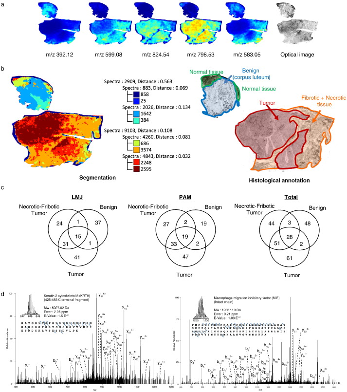
Top-down tissue microproteomics was applied on benign, tumor and necrotic-fibrotic regions of serous ovarian cancer biopsies, identifying proteins exhibiting region-specific cellular localization and PTMs. The regions of interest (ROIs) were determined by MALDI mass spectrometry imaging and spatial segmentation.
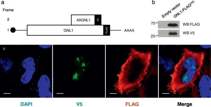
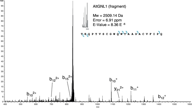
Analysis with a customized protein sequence database containing reference and alternative proteins (altprots) identified 15 altprots, including alternative G protein nucleolar 1 (AltGNL1) found in the tumor, and translated from an altORF nested within the GNL1 canonical coding sequence. Co-expression of GNL1 and altGNL1 was validated by transfection in HEK293 and HeLa cells with an expression plasmid containing a GNL1-FLAG construct. Western blot and immunofluorescence experiments confirmed constitutive co-expression of altGNL1-V5 with GNL1-FLAG.
Taken together, our approach provides means to evaluate protein changes in the case of serous ovarian cancer, allowing the detection of potential markers that have never been considered.
最近,人们已经证明蛋白质可以从替代开放阅读框(altORFs)翻译出来,从而增加实际蛋白质组的大小。基于自上而下的质谱的蛋白质组学允许鉴定含有翻译后修饰(PTMs)的完整蛋白质以及从参考 ORF 或 altORF 翻译而来的截断形式。
在浆液性卵巢癌活检的良性、肿瘤和坏死纤维化区域应用自上而下的组织微蛋白质组学,鉴定表现出区域特异性细胞定位和 PTM 的蛋白质。通过 MALDI 质谱成像和空间分割确定感兴趣区域(ROI)。
使用包含参考和替代蛋白(altprots)的定制蛋白质序列数据库进行分析,鉴定出 15 个 altprots,包括肿瘤中发现的替代 G 蛋白核仁 1(AltGNL1),以及从 GNL1 规范编码序列内嵌套的 altORF 翻译而来。通过用包含 GNL1-FLAG 构建体的表达质粒转染 HEK293 和 HeLa 细胞,验证了 GNL1 和 altGNL1 的共表达。Western blot 和免疫荧光实验证实了 altGNL1-V5 与 GNL1-FLAG 的组成性共表达。
总之,我们的方法提供了评估浆液性卵巢癌情况下蛋白质变化的手段,允许检测以前从未考虑过的潜在标志物。