• 文献检索
  • 文档翻译
  • 深度研究
  • 学术资讯
  • Suppr Zotero 插件Zotero 插件
  • 邀请有礼
  • 套餐&价格
  • 历史记录
应用&插件
Suppr Zotero 插件Zotero 插件浏览器插件Mac 客户端Windows 客户端微信小程序
定价
高级版会员购买积分包购买API积分包
服务
文献检索文档翻译深度研究API 文档MCP 服务
关于我们
关于 Suppr公司介绍联系我们用户协议隐私条款
关注我们

Suppr 超能文献

核心技术专利:CN118964589B侵权必究
粤ICP备2023148730 号-1Suppr @ 2026

文献检索

告别复杂PubMed语法,用中文像聊天一样搜索,搜遍4000万医学文献。AI智能推荐,让科研检索更轻松。

立即免费搜索

文件翻译

保留排版,准确专业,支持PDF/Word/PPT等文件格式,支持 12+语言互译。

免费翻译文档

深度研究

AI帮你快速写综述,25分钟生成高质量综述,智能提取关键信息,辅助科研写作。

立即免费体验

后路松解、内撑开、最终椎弓根椎体截骨术(PSO)及脊柱融合术与一期后路脊柱全椎体切除术治疗多节段重度先天性脊柱侧凸的影像学比较

Radiologic comparison of posterior release, internal distraction, final PSO and spinal fusion with one-stage posterior vertebral column resection for multi-level severe congenital scoliosis.

作者信息

Liu Shichang, Zhang Nannan, Song Yueming, Song Zongrang, Zhang Liping, Liu Jijun, Xie En, Wu Qining, Hao Dingjun

机构信息

Department of Spine Surgery, Honghui Hospital, Xi'an Jiaotong University College of Medicine, South door slightly Friendship Road 555, Xi'an, 710000, People's Republic of China.

National Center for Birth Defect Monitoring, West China Second University Hospital, Sichuan University and Key Laboratory of Birth Defects and Related Diseases of Women and Children (Sichuan University), Ministry of Education, Chengdu, Sichuan, 610041, People's Republic of China.

出版信息

BMC Musculoskelet Disord. 2017 Jun 20;18(1):270. doi: 10.1186/s12891-017-1627-9.

DOI:10.1186/s12891-017-1627-9
PMID:28633648
原文链接:https://pmc.ncbi.nlm.nih.gov/articles/PMC5477743/
Abstract

BACKGROUND

To compare radiologic results of posterior release, internal distraction, and final pedicle subtraction osteotomy (PSO) and spinal fusionwith one-stage posterior vertebral column resection (PVCR) in treating multi-level severe congenital scoliosis.

METHODS

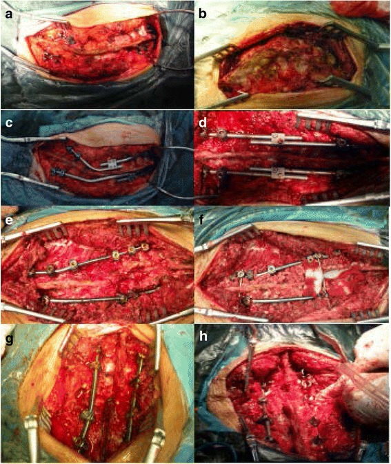
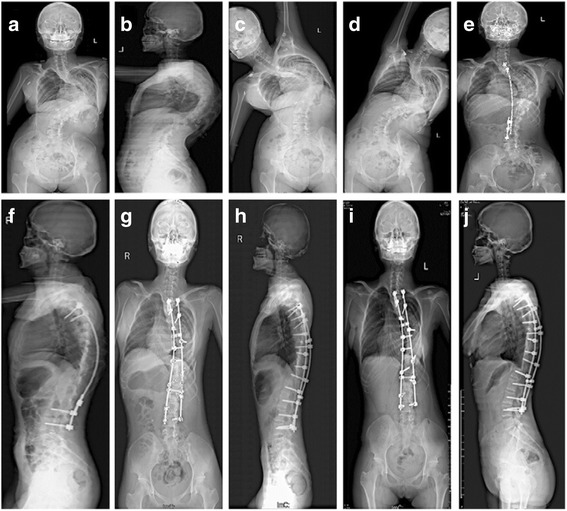
Forty-onesevere congenital scoliosis patients were used in the study. Group A comprised 24 patients who underwent one-stage PVCR. Group B comprised 17 patients who underwent posterior release with internal distraction, followed by final posterior fusion and instrumentation. The average preoperative main curve was 110.4° (95-130°) in group A and 109.4° (range 90°-126°) in group B. Postoperative follow-up time was ≥2 years (2.0-4.5 years) to analyze the radiographic and clinical outcomes.

RESULTS

A comparison of posterior release, internal distraction, and final spinal fusion with PVCR showed no significant differences in postoperative main curve and compensatory caudal curve correction, coronal and sagittal imbalance. However, significant differences were found between the 2 groups in compensatory cranial curve correction.

CONCLUSIONS

Posterior release, internal distraction, and final spinal fusion produce better corrective results in compensatory cranial curve correction than PVCR in treating severe multi-level congenital scoliosis.

摘要

背景

比较后路松解、内撑开以及最终的椎弓根截骨术(PSO)和脊柱融合术与一期后路全脊椎切除术(PVCR)治疗多节段重度先天性脊柱侧凸的影像学结果。

方法

41例重度先天性脊柱侧凸患者纳入本研究。A组24例患者接受一期PVCR。B组17例患者接受后路松解加内撑开,随后行最终的后路融合及内固定术。A组术前平均主弯为110.4°(95 - 130°),B组为109.4°(范围90° - 126°)。术后随访时间≥2年(2.0 - 4.5年),以分析影像学和临床结果。

结果

后路松解、内撑开以及最终的脊柱融合术与PVCR比较,术后主弯及代偿性尾侧弯矫正、冠状面和矢状面失衡方面无显著差异。然而,两组在代偿性头侧弯矫正方面存在显著差异。

结论

在治疗重度多节段先天性脊柱侧凸时,后路松解、内撑开以及最终的脊柱融合术在代偿性头侧弯矫正方面比PVCR产生更好的矫正效果。

https://cdn.ncbi.nlm.nih.gov/pmc/blobs/0400/5477743/1510da22e46e/12891_2017_1627_Fig3_HTML.jpg
https://cdn.ncbi.nlm.nih.gov/pmc/blobs/0400/5477743/6ed6417cf02b/12891_2017_1627_Fig1_HTML.jpg
https://cdn.ncbi.nlm.nih.gov/pmc/blobs/0400/5477743/15a01b141773/12891_2017_1627_Fig2_HTML.jpg
https://cdn.ncbi.nlm.nih.gov/pmc/blobs/0400/5477743/1510da22e46e/12891_2017_1627_Fig3_HTML.jpg
https://cdn.ncbi.nlm.nih.gov/pmc/blobs/0400/5477743/6ed6417cf02b/12891_2017_1627_Fig1_HTML.jpg
https://cdn.ncbi.nlm.nih.gov/pmc/blobs/0400/5477743/15a01b141773/12891_2017_1627_Fig2_HTML.jpg
https://cdn.ncbi.nlm.nih.gov/pmc/blobs/0400/5477743/1510da22e46e/12891_2017_1627_Fig3_HTML.jpg

相似文献

1
Radiologic comparison of posterior release, internal distraction, final PSO and spinal fusion with one-stage posterior vertebral column resection for multi-level severe congenital scoliosis.后路松解、内撑开、最终椎弓根椎体截骨术(PSO)及脊柱融合术与一期后路脊柱全椎体切除术治疗多节段重度先天性脊柱侧凸的影像学比较
BMC Musculoskelet Disord. 2017 Jun 20;18(1):270. doi: 10.1186/s12891-017-1627-9.
2
Congenital scoliosis treated with posterior vertebral column resection in patients younger than 18 years: longer than 10-year follow-up.18岁以下患者采用后路脊柱切除术治疗先天性脊柱侧凸:超过10年的随访
J Neurosurg Spine. 2016 Aug;25(2):225-33. doi: 10.3171/2015.11.SPINE151099. Epub 2016 Mar 11.
3
Anterior release internal distraction and posterior spinal fusion for severe and rigid scoliosis.前路松解内撑开和后路脊柱融合术治疗重度僵硬性脊柱侧凸。
Spine (Phila Pa 1976). 2013 Oct 15;38(22):E1411-7. doi: 10.1097/BRS.0b013e3182a3cd90.
4
Posterior vertebral column resection for correction of rigid spinal deformity curves greater than 100°.后路脊柱截骨术矫正大于 100°的僵硬脊柱畸形曲线。
J Neurosurg Spine. 2012 Dec;17(6):540-51. doi: 10.3171/2012.9.SPINE111026. Epub 2012 Oct 12.
5
Posterior all-pedicle screw instrumentation combined with multiple chevron and concave rib osteotomies in the treatment of adolescent congenital kyphoscoliosis.后路全椎弓根螺钉内固定联合多节段楔形截骨和凹侧楔形截骨矫形术治疗青少年先天性脊柱后凸侧凸畸形。
Spine J. 2014 Jan;14(1):11-9. doi: 10.1016/j.spinee.2012.10.016. Epub 2012 Dec 4.
6
Comparison of anterior and posterior vertebral column resection versus anterior release with posterior internal distraction for severe and rigid scoliosis.严重僵硬型脊柱侧弯前路与后路脊柱切除与前路松解联合后路内撑开术的比较
Eur Spine J. 2014 Jun;23(6):1237-43. doi: 10.1007/s00586-014-3270-6. Epub 2014 Mar 20.
7
Anterior release posterior internal distraction and subsequent posterior spinal fusion for the treatment of severe kyphoscoliosis.前路松解后路内撑开及随后的后路脊柱融合术治疗重度脊柱侧凸。
Eur Spine J. 2015 Jul;24(7):1560-7. doi: 10.1007/s00586-015-3902-5. Epub 2015 Mar 27.
8
Management of sagittal balance in adult spinal deformity with minimally invasive anterolateral lumbar interbody fusion: a preliminary radiographic study.微创前路腰椎椎间融合术治疗成人脊柱畸形的矢状位平衡管理:初步影像学研究。
J Neurosurg Spine. 2014 May;20(5):515-22. doi: 10.3171/2014.2.SPINE1347. Epub 2014 Mar 14.
9
Short-term X-ray results of posterior vertebral column resection in severe congenital kyphosis, scoliosis, and kyphoscoliosis.严重先天性脊柱后凸、脊柱侧凸和后凸脊柱侧凸后路脊柱全长截骨术的短期 X 射线结果。
Spine (Phila Pa 1976). 2012 May 20;37(12):1054-7. doi: 10.1097/BRS.0b013e31823b4142.
10
The impact of posterior temporary internal distraction on stepwise corrective surgery for extremely severe and rigid scoliosis greater than 130°.后路临时内撑开对大于130°的极重度僵硬型脊柱侧凸分步矫正手术的影响
Eur Spine J. 2016 Feb;25(2):557-68. doi: 10.1007/s00586-015-4013-z. Epub 2015 Jun 14.

引用本文的文献

1
A comparison of one-stage posterior hemivertebrectomy and asymmetric osteotomy combined with posterior release for young children with congenital scoliosis.一期后路半椎体切除术与不对称截骨联合后路松解治疗幼儿先天性脊柱侧凸的比较
Eur J Med Res. 2025 Jun 10;30(1):470. doi: 10.1186/s40001-025-02732-y.
2
Surgical outcome of scoliosis in patients with Marfan syndrome.马凡综合征患者脊柱侧凸的手术治疗效果。
Spine Deform. 2022 Nov;10(6):1453-1460. doi: 10.1007/s43390-022-00547-z. Epub 2022 Jul 30.
3
Relationship between BMP2/9 Levels and Spinal Function and Quality of Life in Patients with Severe Scoliosis after PVCR.

本文引用的文献

1
Etiopathogenesis of adolescent idiopathic scoliosis: Expression of melatonin receptors 1A/1B, calmodulin and estrogen receptor 2 in deep paravertebral muscles revisited.青少年特发性脊柱侧凸的病因发病机制:重新审视褪黑素受体1A/1B、钙调蛋白和雌激素受体2在椎旁深层肌肉中的表达
Mol Med Rep. 2016 Dec;14(6):5719-5724. doi: 10.3892/mmr.2016.5927. Epub 2016 Nov 7.
2
Understanding the role of the immune system in adolescent idiopathic scoliosis: Immunometabolic CONnections to Scoliosis (ICONS) study protocol.了解免疫系统在青少年特发性脊柱侧凸中的作用:脊柱侧凸的免疫代谢关联(ICONS)研究方案。
BMJ Open. 2016 Jul 8;6(7):e011812. doi: 10.1136/bmjopen-2016-011812.
3
PVCR术后重度脊柱侧弯患者BMP2/9水平与脊柱功能及生活质量的关系
Evid Based Complement Alternat Med. 2021 Oct 29;2021:7008898. doi: 10.1155/2021/7008898. eCollection 2021.
4
Transpedicular bi-vertebrae wedge osteotomy in treatment of post-tubercular spinal deformity: a retrospective study.经椎弓根双椎体楔形截骨术治疗结核后脊柱畸形:一项回顾性研究
BMC Musculoskelet Disord. 2021 Apr 12;22(1):345. doi: 10.1186/s12891-021-04220-w.
5
Vertebral column decancellation in Pott's deformity: use of Surgimap Spine for preoperative surgical planning, retrospective review of 18 patients.脊柱结核畸形中的椎体去松质骨术:使用Surgimap Spine进行术前手术规划,对18例患者的回顾性研究
BMC Musculoskelet Disord. 2018 Jan 15;19(1):13. doi: 10.1186/s12891-018-1929-6.
Congenital scoliosis treated with posterior vertebral column resection in patients younger than 18 years: longer than 10-year follow-up.
18岁以下患者采用后路脊柱切除术治疗先天性脊柱侧凸:超过10年的随访
J Neurosurg Spine. 2016 Aug;25(2):225-33. doi: 10.3171/2015.11.SPINE151099. Epub 2016 Mar 11.
4
One-stage posterior osteotomy with short segmental fusion and dual growing rod technique for severe rigid congenital scoliosis: the preliminary clinical outcomes of a hybrid technique.一期后路截骨短节段融合及双生长棒技术治疗重度僵硬型先天性脊柱侧凸:一种混合技术的初步临床疗效
Spine (Phila Pa 1976). 2014 Feb 15;39(4):E294-9. doi: 10.1097/BRS.0000000000000119.
5
The risk factors of neurologic deficits of one-stage posterior vertebral column resection for patients with severe and rigid spinal deformities.重度僵硬脊柱畸形患者一期后路脊柱全切除术后神经功能缺损的危险因素。
Eur Spine J. 2014 Jan;23(1):149-56. doi: 10.1007/s00586-013-2793-6. Epub 2013 Apr 26.
6
Dual growing rods technique for congenital scoliosis: more than 2 years outcomes: preliminary results of a single center.双生长棒技术治疗先天性脊柱侧凸:超过 2 年的结果:单中心初步结果。
Spine (Phila Pa 1976). 2012 Dec 15;37(26):E1639-44. doi: 10.1097/BRS.0b013e318273d6bf.
7
Posterior extrapleural intervertebral space release combined with wedge osteotomy for the treatment of severe rigid scoliosis.后路胸膜外椎间松解联合楔形截骨术治疗重度僵硬性脊柱侧凸。
Spine (Phila Pa 1976). 2012 May 15;37(11):E647-54. doi: 10.1097/BRS.0b013e318250042b.
8
Surgical treatment of severe scoliosis and kyphoscoliosis by stages.分期手术治疗重度脊柱侧凸和后凸畸形
Chin Med J (Engl). 2012 Jan;125(1):81-6.
9
Short-term X-ray results of posterior vertebral column resection in severe congenital kyphosis, scoliosis, and kyphoscoliosis.严重先天性脊柱后凸、脊柱侧凸和后凸脊柱侧凸后路脊柱全长截骨术的短期 X 射线结果。
Spine (Phila Pa 1976). 2012 May 20;37(12):1054-7. doi: 10.1097/BRS.0b013e31823b4142.
10
Anterior and posterior vertebral column resection for severe and rigid idiopathic scoliosis.脊柱前路和后路截骨术治疗重度僵硬特发性脊柱侧凸。
Eur Spine J. 2011 Oct;20(10):1728-34. doi: 10.1007/s00586-011-1861-z. Epub 2011 Jun 2.