Wu Ling-Yun, Ye Zhen-Nan, Zhou Chen-Hui, Wang Chun-Xi, Xie Guang-Bin, Zhang Xiang-Sheng, Gao Yong-Yue, Zhang Zi-Huan, Zhou Meng-Liang, Zhuang Zong, Liu Jing-Peng, Hang Chun-Hua, Shi Ji-Xin
Department of Neurosurgery, Jinling Hospital, School of Medicine, Nanjing UniversityNanjing, China.
Department of Neurosurgery, The Second Affiliated Hospital of Guangzhou Medical UniversityGuangzhou, China.
Front Mol Neurosci. 2017 Jun 6;10:175. doi: 10.3389/fnmol.2017.00175. eCollection 2017.
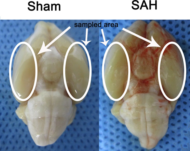
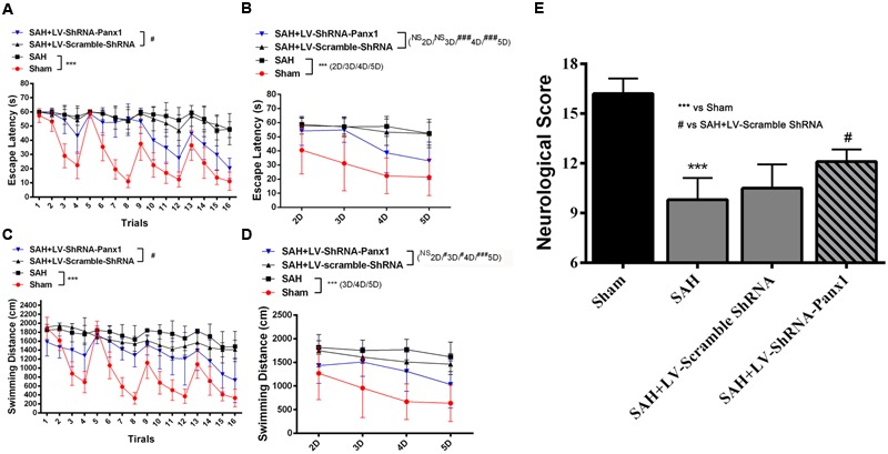
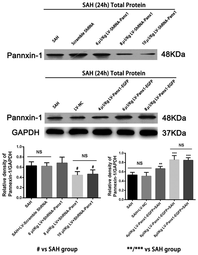
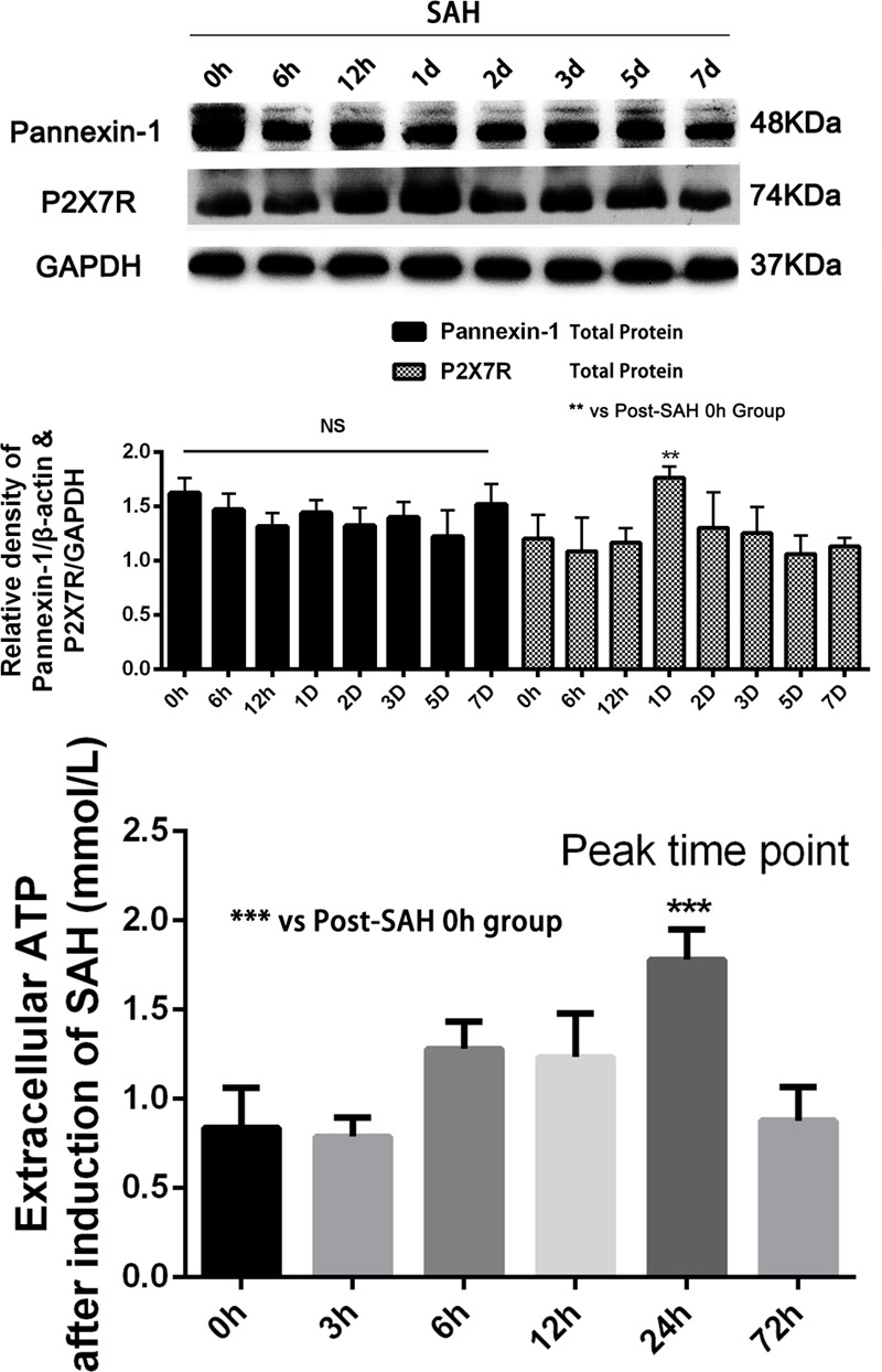
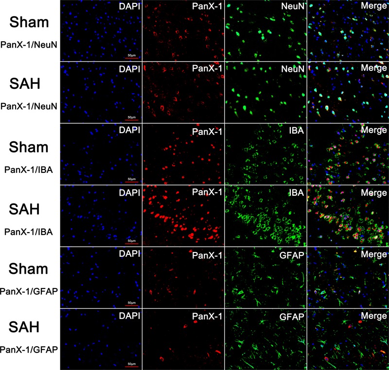
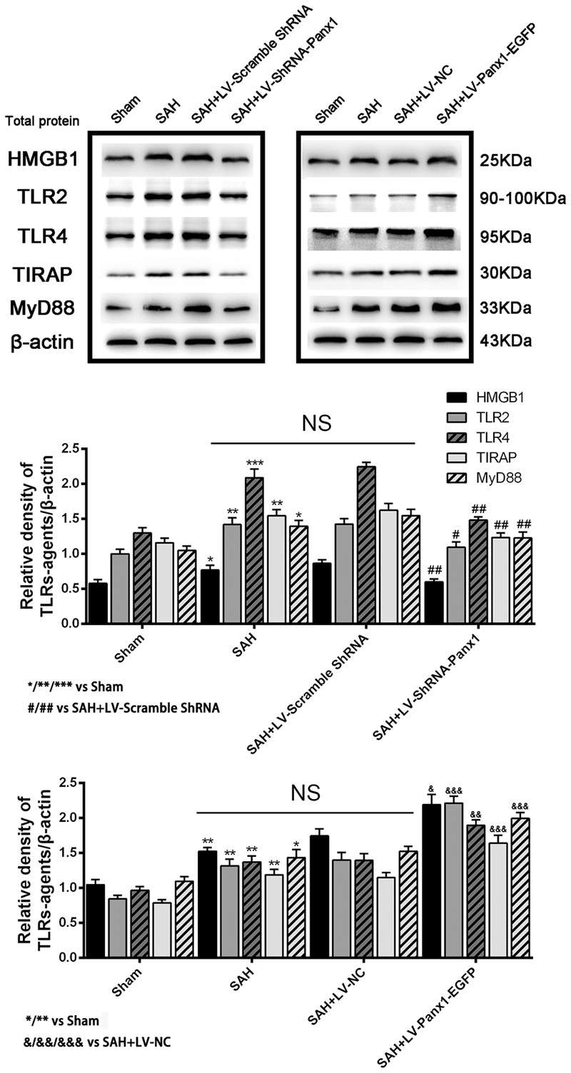
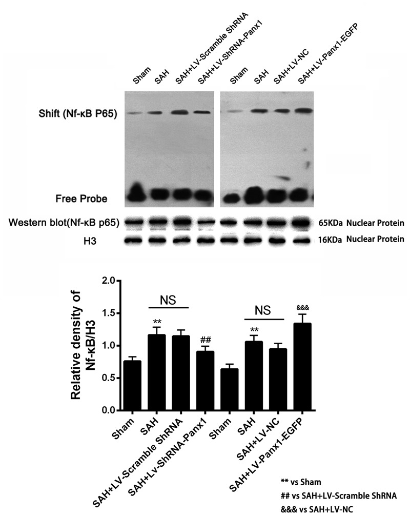
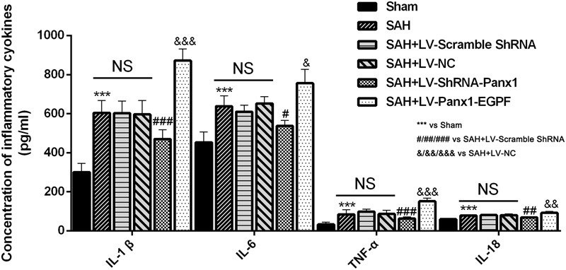
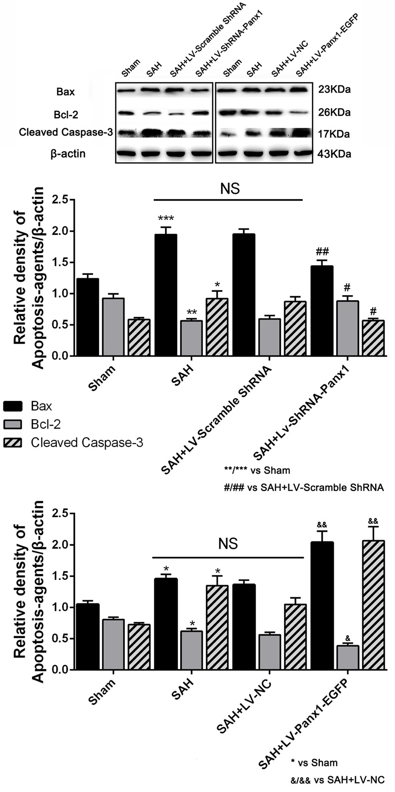
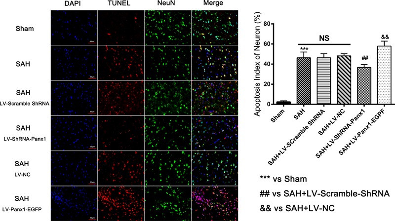
Accumulating evidence suggests that neuroinflammation plays a critical role in early brain injury after subarachnoid hemorrhage (SAH). Pannexin-1 channels, as a member of gap junction proteins located on the plasma membrane, releases ATP, ions, second messengers, neurotransmitters, and molecules up to 1 kD into the extracellular space, when activated. Previous studies identified that the opening of Pannexin-1 channels is essential for cellular migration, apoptosis and especially inflammation, but its effects on inflammatory response in SAH model have not been explored yet. Adult male Sprague-Dawley rats were divided into six groups: sham group ( = 20), SAH group ( = 20), SAH + LV-Scramble-ShRNA group ( = 20), SAH + LV-ShRNA-Panx1 group ( = 20), SAH + LV-NC group ( = 20), and SAH + LV-Panx1-EGFP group ( = 20). The rat SAH model was induced by injection of 0.3 ml fresh arterial, non-heparinized blood into the prechiasmatic cistern in 20 s. In SAH + LV-ShRNA-Panx1 group and SAH + LV-Panx1-EGFP group, lentivirus was administered via intracerebroventricular injection (i.c.v.) at 72 h before the induction of SAH. The Quantitative real-time polymerase chain reaction, electrophoretic mobility shift assay, enzyme-linked immunosorbent assay, immunofluorescence staining, and western blotting were performed to explore the potential interactive mechanism between Pannexin-1 channels and TLR2/TLR4/NF-κB-mediated signaling pathway. Cognitive and memory changes were investigated by the Morris water maze test. Administration with LV-ShRNA-Panx1 markedly decreased the expression levels of TLR2/4/NF-κB pathway-related agents in the brain cortex and significantly ameliorated neurological cognitive and memory deficits in this SAH model. On the contrary, administration of LV-Panx1-EGFP elevated the expressions of TLR2/4/NF-κB pathway-related agents, which correlated with augmented neuronal apoptosis. Pannexin-1 channels may contribute to inflammatory response and neurobehavioral dysfunction through the TLR2/TLR4/NF-κB-mediated pathway signaling after SAH, suggesting a potential role of Pannexin-1 channels could be a potential therapeutic target for the treatment of SAH.
越来越多的证据表明,神经炎症在蛛网膜下腔出血(SAH)后的早期脑损伤中起关键作用。泛素蛋白-1通道作为位于质膜上的间隙连接蛋白成员,在被激活时会将三磷酸腺苷(ATP)、离子、第二信使、神经递质以及分子量高达1千道尔顿的分子释放到细胞外空间。先前的研究表明,泛素蛋白-1通道的开放对细胞迁移、凋亡尤其是炎症至关重要,但尚未探究其在SAH模型中对炎症反应的影响。成年雄性Sprague-Dawley大鼠被分为六组:假手术组(n = 20)、SAH组(n = 20)、SAH + LV-乱序短发夹RNA组(n = 20)、SAH + LV-泛素蛋白-1短发夹RNA组(n = 20)、SAH + LV-阴性对照(NC)组(n = 20)以及SAH + LV-泛素蛋白-1-增强绿色荧光蛋白(EGFP)组(n = 20)。通过在20秒内向视交叉前池注射0.3毫升新鲜动脉非肝素化血液诱导大鼠SAH模型。在SAH + LV-泛素蛋白-1短发夹RNA组和SAH + LV-泛素蛋白-1-EGFP组中,在诱导SAH前72小时通过脑室内注射(i.c.v.)给予慢病毒。采用定量实时聚合酶链反应、电泳迁移率变动分析、酶联免疫吸附测定、免疫荧光染色和蛋白质印迹法来探究泛素蛋白-1通道与Toll样受体2(TLR2)/Toll样受体4(TLR4)/核因子κB(NF-κB)介导的信号通路之间潜在的相互作用机制。通过莫里斯水迷宫试验研究认知和记忆变化。给予LV-泛素蛋白-1短发夹RNA显著降低了大脑皮质中TLR2/4/NF-κB通路相关因子的表达水平,并显著改善了该SAH模型中的神经认知和记忆缺陷。相反,给予LV-泛素蛋白-EGFP提高了TLR2/4/NF-κB通路相关因子的表达,这与神经元凋亡增加相关。SAH后,泛素蛋白-1通道可能通过TLR2/TLR4/NF-κB介导的信号通路促进炎症反应和神经行为功能障碍,这表明泛素蛋白-1通道可能是治疗SAH的潜在治疗靶点。