Darley David R, Granger Emily, Glanville Allan R
Department of Thoracic Medicine St Vincent's Hospital Darlinghurst NSW Australia.
Department of Cardiothoracic Surgery St Vincent's Hospital Darlinghurst NSW Australia.
Respirol Case Rep. 2017 Jun 15;5(5):e00250. doi: 10.1002/rcr2.250. eCollection 2017 Sep.
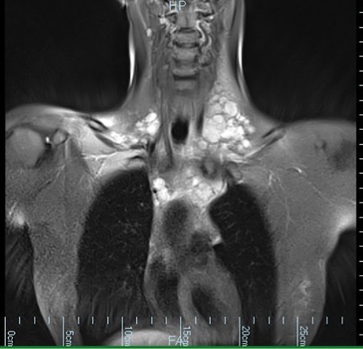
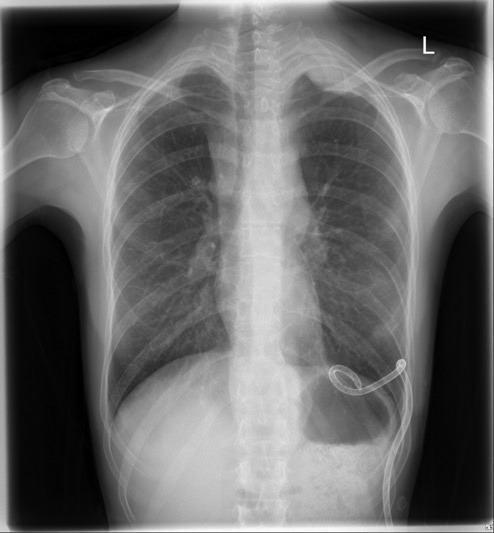
Malignant mesothelioma presenting with recurrent chylous effusion is rare. We describe the case of a 34-year-old female Macedonian immigrant who presented with central chest pain and subsequently a left-sided chylous pleural effusion. The diagnosis was made on pleural biopsy via video-assisted thoracoscopic surgery (VATS). Our case demonstrates the utility of thoracic magnetic resonance imaging (MRI) and the difficulties associated with pleural cytology and cervical lymph node biopsy in the establishment of a diagnosis of mesothelioma. It is a reminder that mesothelioma can metastasize to mediastinal and cervical lymph nodes, can occur in young people, and may present as a chylothorax.
以复发性乳糜性胸腔积液为表现的恶性间皮瘤较为罕见。我们报告一例34岁的马其顿女性移民病例,该患者最初表现为胸痛,随后出现左侧乳糜性胸腔积液。诊断通过电视辅助胸腔镜手术(VATS)进行胸膜活检得以确立。我们的病例展示了胸部磁共振成像(MRI)的作用,以及在间皮瘤诊断过程中胸膜细胞学检查和颈部淋巴结活检所面临的困难。这提醒我们,间皮瘤可转移至纵隔和颈部淋巴结,可发生于年轻人,且可能以乳糜胸的形式出现。