Assistance Publique - Hôpitaux de Paris (AP-HP), Hôpital Saint-Antoine, Service de réanimation médicale, 184 rue du Faubourg Saint-Antoine, 75571, Paris, Cedex 12, France.
Université Pierre-et-Marie Curie, Paris 6, France.
Crit Care. 2017 Jun 23;21(1):155. doi: 10.1186/s13054-017-1742-x.
Mottling around the knee, reflecting a reduced skin blood flow, is predictive of mortality in patients with septic shock. However, the causative pathophysiology of mottling remains unknown. We hypothesized that the cutaneous hypoperfusion observed in the mottled area is related to regional endothelial dysfunction.
This was a prospective, observational study in a medical ICU in a tertiary teaching hospital. Consecutive adult patients with sepsis admitted to ICU were included. After resuscitation, endothelium-dependent vasodilation in the skin circulation was measured before and after iontophoresis of acetylcholine (Ach) in the forearm and the knee area. We analyzed the patterns of induced vasodilatation according to the presence or absence of mottling and vital status at 14 days.
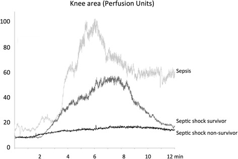
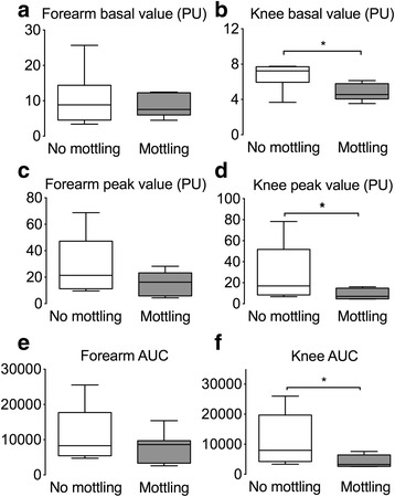
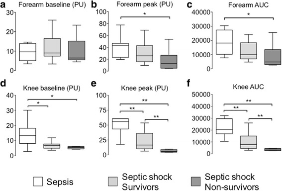
We evaluated 37 septic patients, including 11 without and 26 with septic shock. Overall 14-day mortality was 22%. Ten patients had mottling around the knee (10/37, 27%). In the knee area, the increased skin blood flow following iontophoresis of Ach was lower in patients with mottled skin as compared to patients without mottled skin (area under curve (AUC) 3280 (2643-6440) vs. 7980 (4233-19,707), both P < 0.05). In the forearm area, the increased skin blood flow following iontophoresis of Ach was similar in patients with and without mottled skin. Among patients with septic shock, the increased skin blood flow following iontophoresis of Ach in the knee area was significantly lower in non-survivors as compared to survivors at 14 days (AUC 3256 (2600-4426) vs. 7704 (4539-15,011), P < 0.01). In patients with septic shock, the increased skin blood flow in the forearm area following iontophoresis of Ach was similar in survivors and non-survivors at 14 days.
Mottling is associated with regional endothelial dysfunction in patients with septic shock. Endothelial dysfunction in the knee skin area was more pronounced in non-survivors than in survivors.
膝关节周围出现斑点,反映皮肤血流量减少,可预测感染性休克患者的死亡率。然而,斑点形成的病理生理学机制尚不清楚。我们假设观察到的斑点区域皮肤灌注不足与局部内皮功能障碍有关。
这是一项在一家三级教学医院的重症监护病房进行的前瞻性观察性研究。纳入因败血症入住 ICU 的成年患者。在复苏后,在前臂和膝关节区域进行乙酰胆碱(Ach)离子电渗后,测量皮肤循环的内皮依赖性血管扩张。我们根据斑点的有无和 14 天的生存状态分析诱导血管扩张的模式。
我们评估了 37 例败血症患者,其中 11 例无休克,26 例休克。总体 14 天死亡率为 22%。10 例患者膝关节周围有斑点(10/37,27%)。与无斑点皮肤的患者相比,接受 Ach 离子电渗后膝关节皮肤血流增加量较低(曲线下面积(AUC)3280(2643-6440)vs. 7980(4233-19707),均 P<0.05)。在前臂区域,接受 Ach 离子电渗后皮肤血流增加量在有斑点和无斑点皮肤的患者中相似。在感染性休克患者中,与存活者相比,14 天时接受 Ach 离子电渗后膝关节皮肤血流增加量在非存活者中显著降低(AUC 3256(2600-4426)vs. 7704(4539-15011),P<0.01)。在感染性休克患者中,与存活者相比,14 天时接受 Ach 离子电渗后前臂皮肤血流增加量在非存活者中相似。
斑点形成与感染性休克患者的局部内皮功能障碍有关。与存活者相比,非存活者膝关节皮肤区域的内皮功能障碍更为明显。