• 文献检索
  • 文档翻译
  • 深度研究
  • 学术资讯
  • Suppr Zotero 插件Zotero 插件
  • 邀请有礼
  • 套餐&价格
  • 历史记录
应用&插件
Suppr Zotero 插件Zotero 插件浏览器插件Mac 客户端Windows 客户端微信小程序
定价
高级版会员购买积分包购买API积分包
服务
文献检索文档翻译深度研究API 文档MCP 服务
关于我们
关于 Suppr公司介绍联系我们用户协议隐私条款
关注我们

Suppr 超能文献

核心技术专利:CN118964589B侵权必究
粤ICP备2023148730 号-1Suppr @ 2026

文献检索

告别复杂PubMed语法,用中文像聊天一样搜索,搜遍4000万医学文献。AI智能推荐,让科研检索更轻松。

立即免费搜索

文件翻译

保留排版,准确专业,支持PDF/Word/PPT等文件格式,支持 12+语言互译。

免费翻译文档

深度研究

AI帮你快速写综述,25分钟生成高质量综述,智能提取关键信息,辅助科研写作。

立即免费体验

晚期非小细胞肺癌氟-氟莫司汀摄取:体素 PET 动力学研究。

F-fluoromisonidazole uptake in advanced stage non-small cell lung cancer: A voxel-by-voxel PET kinetics study.

机构信息

Cancer Research UK/MRC Oxford Institute for Radiation Oncology, Gray Laboratories, Department of Oncology, University of Oxford, Oxford, UK.

Radiation Physics and Protection, Oxford University Hospitals NHS Foundation Trust, Oxford, UK.

出版信息

Med Phys. 2017 Sep;44(9):4665-4676. doi: 10.1002/mp.12416. Epub 2017 Jul 21.

DOI:10.1002/mp.12416
PMID:28644546
原文链接:https://pmc.ncbi.nlm.nih.gov/articles/PMC5600259/
Abstract

PURPOSE

The aim of this study was to determine the relative abilities of compartment models to describe time-courses of F-fluoromisonidazole (FMISO) uptake in tumor voxels of patients with non-small cell lung cancer (NSCLC) imaged using dynamic positron emission tomography. Also to use fits of the best-performing model to investigate changes in fitted rate-constants with distance from the tumor edge.

METHODS

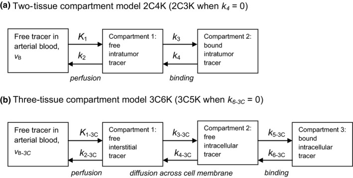
Reversible and irreversible two- and three-tissue compartment models were fitted to 24 662 individual voxel time activity curves (TACs) obtained from tumors in nine patients, each imaged twice. Descriptions of the TACs provided by the models were compared using the Akaike and Bayesian information criteria (AIC and BIC). Two different models (two- and three-tissue) were fitted to 30 measured voxel TACs to provide ground-truth TACs for a statistical simulation study. Appropriately scaled noise was added to each of the resulting ground-truth TACs, generating 1000 simulated noisy TACs for each ground-truth TAC. The simulation study was carried out to provide estimates of the accuracy and precision with which parameter values are determined, the estimates being obtained for both assumptions about the ground-truth kinetics. A BIC clustering technique was used to group the fitted rate-constants, taking into consideration the underlying uncertainties on the fitted rate-constants. Voxels were also categorized according to their distance from the tumor edge.

RESULTS

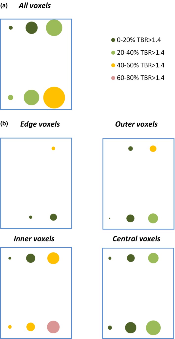
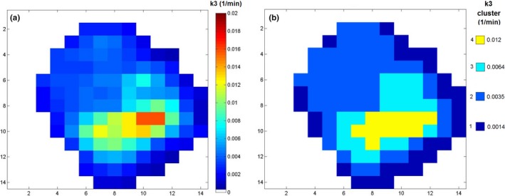
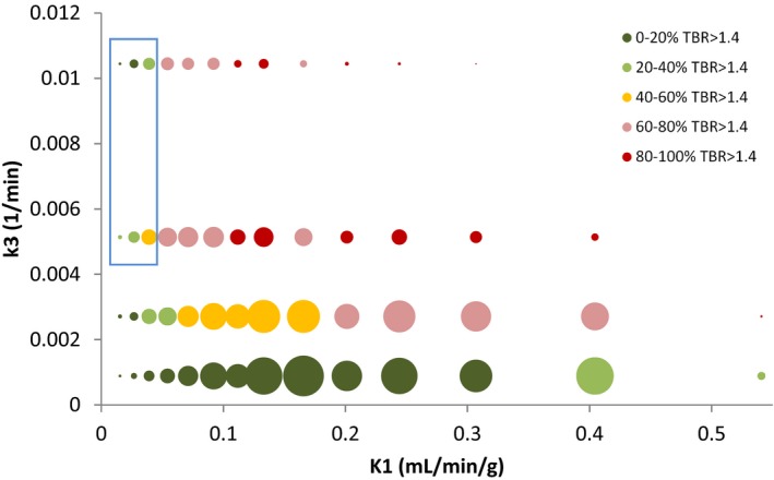
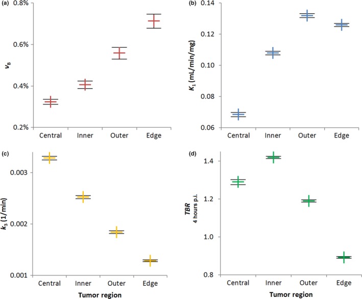
For uptake time-courses of individual voxels an irreversible two-tissue compartment model was found to be most precise. The simulation study indicated that this model had a one standard deviation precision of 39% for tumor fractional blood volumes and 37% for the FMISO binding rate-constant. Weighted means of fitted FMISO binding rate-constants of voxels in all tumors rose significantly with increasing distance from the tumor edge, whereas fitted fractional blood volumes fell significantly. When grouped using the BIC clustering, many centrally located voxels had high-fitted FMISO binding rate-constants and low rate-constants for tracer flow between the vasculature and tumor, both indicative of hypoxia. Nevertheless, many of these voxels had tumor-to-blood (TBR) values lower than the 1.4 level commonly expected for hypoxic tissues, possibly due to the low rate-constants for tracer flow between the vasculature and tumor cells in these voxels.

CONCLUSIONS

Time-courses of FMISO uptake in NSCLC tumor voxels are best analyzed using an irreversible two-tissue compartment model, fits of which provide more precise parameter values than those of a three-tissue model. Changes in fitted model parameter values indicate that levels of hypoxia rise with increasing distance from tumor edges. The average FMISO binding rate-constant is higher for voxels in tumor centers than in the next tumor layer out, but the average value of the more simplistic TBR metric is lower in tumor centers. For both metrics, higher values might be considered indicative of hypoxia, and the mismatch in this case is likely to be due to poor perfusion at the tumor center. Kinetics analysis of dynamic PET images may therefore provide more accurate measures of the hypoxic status of such regions than the simpler TBR metric, a hypothesis we are presently exploring in a study of tumor imaging versus histopathology.

摘要

目的

本研究旨在确定房室模型相对能力,以描述使用动态正电子发射断层扫描对非小细胞肺癌(NSCLC)患者肿瘤体素进行氟代米索硝唑(FMISO)摄取的时间过程。还利用表现最佳模型的拟合来研究肿瘤边缘距离与拟合率常数的变化。

方法

将可逆和不可逆的两室和三室房室模型拟合到 9 名患者的 24662 个肿瘤体素时间活动曲线(TAC),每个患者都进行了两次成像。使用 Akaike 和贝叶斯信息准则(AIC 和 BIC)比较模型对 TAC 的描述。将两种不同的模型(两室和三室)拟合到 30 个测量的体素 TAC,为统计模拟研究提供地面真实 TAC。为每个地面真实 TAC 适当缩放噪声,为每个地面真实 TAC 生成 1000 个模拟噪声 TAC。进行了模拟研究,以提供参数值确定的准确性和精密度的估计值,这两个估计值都是基于对地面真实动力学的假设。使用 BIC 聚类技术根据拟合率常数的潜在不确定性对其进行分组。还根据与肿瘤边缘的距离对体素进行分类。

结果

对于个体体素的摄取时间过程,发现不可逆的两室房室模型最为精确。模拟研究表明,对于肿瘤的部分血容量,该模型的标准差精度为 39%,对于 FMISO 结合率常数的标准差精度为 37%。所有肿瘤中体素拟合的 FMISO 结合率常数的加权平均值随着与肿瘤边缘距离的增加而显著升高,而拟合的部分血容量则显著降低。使用 BIC 聚类进行分组时,许多中央体素具有高拟合的 FMISO 结合率常数和示踪剂在脉管系统和肿瘤之间流动的低率常数,这两者都表明存在缺氧。尽管如此,许多这些体素的肿瘤与血液(TBR)值低于 1.4,这是缺氧组织通常预期的水平,这可能是由于这些体素中示踪剂在脉管系统和肿瘤细胞之间的流动率常数较低。

结论

使用不可逆的两室房室模型对 NSCLC 肿瘤体素的 FMISO 摄取时间过程进行分析最佳,其拟合提供了比三室模型更精确的参数值。拟合模型参数值的变化表明,缺氧水平随肿瘤边缘距离的增加而升高。位于肿瘤中心的体素的平均 FMISO 结合率常数高于下一肿瘤层的体素,但肿瘤中心的更简单 TBR 指标的平均值较低。对于这两个指标,较高的值可能被认为是缺氧的指标,而在这种情况下的不匹配很可能是由于肿瘤中心的灌注不良。因此,与简单的 TBR 指标相比,动态 PET 图像的动力学分析可能为这些区域的缺氧状态提供更准确的测量,我们目前正在一项肿瘤成像与组织病理学的研究中探索这一假设。

https://cdn.ncbi.nlm.nih.gov/pmc/blobs/b32e/5638114/7d17ad1986ab/MP-44-4665-g007.jpg
https://cdn.ncbi.nlm.nih.gov/pmc/blobs/b32e/5638114/0da3d2108472/MP-44-4665-g001.jpg
https://cdn.ncbi.nlm.nih.gov/pmc/blobs/b32e/5638114/8b8ee1382667/MP-44-4665-g002.jpg
https://cdn.ncbi.nlm.nih.gov/pmc/blobs/b32e/5638114/e39c1bf8371b/MP-44-4665-g003.jpg
https://cdn.ncbi.nlm.nih.gov/pmc/blobs/b32e/5638114/ccf0f2566eff/MP-44-4665-g004.jpg
https://cdn.ncbi.nlm.nih.gov/pmc/blobs/b32e/5638114/91328924dd3f/MP-44-4665-g005.jpg
https://cdn.ncbi.nlm.nih.gov/pmc/blobs/b32e/5638114/2f1de50d72cd/MP-44-4665-g006.jpg
https://cdn.ncbi.nlm.nih.gov/pmc/blobs/b32e/5638114/7d17ad1986ab/MP-44-4665-g007.jpg

相似文献

1
F-fluoromisonidazole uptake in advanced stage non-small cell lung cancer: A voxel-by-voxel PET kinetics study.晚期非小细胞肺癌氟-氟莫司汀摄取:体素 PET 动力学研究。
Med Phys. 2017 Sep;44(9):4665-4676. doi: 10.1002/mp.12416. Epub 2017 Jul 21.
2
Whole tumor kinetics analysis of F-fluoromisonidazole dynamic PET scans of non-small cell lung cancer patients, and correlations with perfusion CT blood flow.非小细胞肺癌患者F-氟米索硝唑动态PET扫描的全肿瘤动力学分析及其与灌注CT血流的相关性。
EJNMMI Res. 2018 Aug 1;8(1):73. doi: 10.1186/s13550-018-0430-4.
3
18F-FLT uptake kinetics in head and neck squamous cell carcinoma: a PET imaging study.18F-FLT摄取动力学在头颈部鳞状细胞癌中的研究:一项PET成像研究
Med Phys. 2014 Apr;41(4):041911. doi: 10.1118/1.4868462.
4
Pharmacokinetic Analysis of Dynamic F-Fluoromisonidazole PET Data in Non-Small Cell Lung Cancer.非小细胞肺癌中动态F-氟米索硝唑PET数据的药代动力学分析
J Nucl Med. 2017 Jun;58(6):911-919. doi: 10.2967/jnumed.116.180422. Epub 2017 Feb 23.
5
Reproducibility of intratumor distribution of (18)F-fluoromisonidazole in head and neck cancer.(18)F-氟米索硝唑在头颈部肿瘤内分布的可重复性
Int J Radiat Oncol Biol Phys. 2008 Jan 1;70(1):235-42. doi: 10.1016/j.ijrobp.2007.08.036.
6
Quantification of Tumor Hypoxic Fractions Using Positron Emission Tomography with [F]Fluoromisonidazole ([F]FMISO) Kinetic Analysis and Invasive Oxygen Measurements.利用正电子发射断层扫描结合 [F]氟米索硝唑([F]FMISO)动力学分析和侵袭性氧测量对肿瘤乏氧分数进行定量。
Mol Imaging Biol. 2017 Dec;19(6):893-902. doi: 10.1007/s11307-017-1083-9.
7
F-Fluoromisonidazole Kinetic Modeling for Characterization of Tumor Perfusion and Hypoxia in Response to Antiangiogenic Therapy.用于表征抗血管生成治疗反应中肿瘤灌注和缺氧的F-氟米索硝唑动力学建模
J Nucl Med. 2017 Oct;58(10):1567-1573. doi: 10.2967/jnumed.117.190892. Epub 2017 Mar 30.
8
Interobserver agreement of qualitative analysis and tumor delineation of 18F-fluoromisonidazole and 3'-deoxy-3'-18F-fluorothymidine PET images in lung cancer.肺癌中 18F-氟咪索硝唑和 3'-去氧-3'-18F-氟胸腺嘧啶 PET 图像的定性分析和肿瘤勾画的观察者间一致性。
J Nucl Med. 2013 Sep;54(9):1543-50. doi: 10.2967/jnumed.112.118083. Epub 2013 Aug 5.
9
Timing of hypoxia PET/CT imaging after 18F-fluoromisonidazole injection in non-small cell lung cancer patients.非小细胞肺癌患者注射 18F-氟咪索硝唑后行缺氧正电子发射断层扫描/计算机断层扫描成像的时机。
Sci Rep. 2022 Dec 16;12(1):21746. doi: 10.1038/s41598-022-26199-7.
10
Comparison of Hypermetabolic and Hypoxic Volumes Delineated on [F]FDG and [F]Fluoromisonidazole PET/CT in Non-small-cell Lung Cancer Patients.比较[F]FDG 和 [F]氟米索硝唑 PET/CT 勾画的非小细胞肺癌患者高代谢和低氧体积。
Mol Imaging Biol. 2020 Jun;22(3):764-771. doi: 10.1007/s11307-019-01422-6.

引用本文的文献

1
Evaluations of an Early Change in Tumor Pathophysiology in Response to Radiotherapy with Oxygen Enhanced Electron Paramagnetic Resonance Imaging (OE EPRI).用氧增强电子顺磁共振成像(OE EPRI)评估放疗中肿瘤病理生理学的早期变化。
Mol Imaging Biol. 2024 Jun;26(3):448-458. doi: 10.1007/s11307-024-01925-x. Epub 2024 Jun 13.
2
Timing of hypoxia PET/CT imaging after 18F-fluoromisonidazole injection in non-small cell lung cancer patients.非小细胞肺癌患者注射 18F-氟咪索硝唑后行缺氧正电子发射断层扫描/计算机断层扫描成像的时机。
Sci Rep. 2022 Dec 16;12(1):21746. doi: 10.1038/s41598-022-26199-7.
3
Radiogenomic analysis of primary breast cancer reveals [18F]-fluorodeoxglucose dynamic flux-constants are positively associated with immune pathways and outperform static uptake measures in associating with glucose metabolism.

本文引用的文献

1
Kinetic modeling in PET imaging of hypoxia.缺氧的正电子发射断层显像(PET)成像中的动力学建模
Am J Nucl Med Mol Imaging. 2014 Sep 6;4(6):490-506. eCollection 2014.
2
Interstitial diffusion and the relationship between compartment modelling and multi-scale spatial-temporal modelling of (18)F-FLT tumour uptake dynamics.(18)F-FLT肿瘤摄取动力学的间质扩散以及房室模型与多尺度时空模型之间的关系
Phys Med Biol. 2014 Sep 7;59(17):5175-202. doi: 10.1088/0031-9155/59/17/5175. Epub 2014 Aug 20.
3
18F-FLT uptake kinetics in head and neck squamous cell carcinoma: a PET imaging study.
原发性乳腺癌的放射基因组分析显示,[18F]-氟脱氧葡萄糖的动态通量常数与免疫途径呈正相关,并且在与葡萄糖代谢相关方面优于静态摄取测量。
Breast Cancer Res. 2022 May 17;24(1):34. doi: 10.1186/s13058-022-01529-9.
4
Investigation of atovaquone-induced spatial changes in tumour hypoxia assessed by hypoxia PET/CT in non-small cell lung cancer patients.通过缺氧PET/CT评估非小细胞肺癌患者中阿托伐醌诱导的肿瘤缺氧空间变化的研究。
EJNMMI Res. 2021 Dec 29;11(1):130. doi: 10.1186/s13550-021-00871-x.
5
Elimination of tumor hypoxia by eribulin demonstrated by F-FMISO hypoxia imaging in human tumor xenograft models.在人肿瘤异种移植模型中,通过F-FMISO缺氧成像证明了艾日布林可消除肿瘤缺氧。
EJNMMI Res. 2019 Jun 3;9(1):51. doi: 10.1186/s13550-019-0521-x.
6
How rapid advances in imaging are defining the future of precision radiation oncology.影像学的快速发展如何定义精准放射肿瘤学的未来。
Br J Cancer. 2019 Apr;120(8):779-790. doi: 10.1038/s41416-019-0412-y. Epub 2019 Mar 26.
7
Longitudinal PET imaging of tumor hypoxia during the course of radiotherapy.放疗过程中肿瘤乏氧的纵向 PET 成像。
Eur J Nucl Med Mol Imaging. 2018 Nov;45(12):2201-2217. doi: 10.1007/s00259-018-4116-y. Epub 2018 Aug 20.
8
High Single Doses of Radiation May Induce Elevated Levels of Hypoxia in Early-Stage Non-Small Cell Lung Cancer Tumors.高单次剂量辐射可能会导致早期非小细胞肺癌肿瘤缺氧水平升高。
Int J Radiat Oncol Biol Phys. 2018 Sep 1;102(1):174-183. doi: 10.1016/j.ijrobp.2018.05.032. Epub 2018 Jun 1.
9
Whole tumor kinetics analysis of F-fluoromisonidazole dynamic PET scans of non-small cell lung cancer patients, and correlations with perfusion CT blood flow.非小细胞肺癌患者F-氟米索硝唑动态PET扫描的全肿瘤动力学分析及其与灌注CT血流的相关性。
EJNMMI Res. 2018 Aug 1;8(1):73. doi: 10.1186/s13550-018-0430-4.
10
4D-PET reconstruction using a spline-residue model with spatial and temporal roughness penalties.基于时空粗糙度惩罚的样条残差模型的 4D-PET 重建。
Phys Med Biol. 2018 May 4;63(9):095013. doi: 10.1088/1361-6560/aabb62.
18F-FLT摄取动力学在头颈部鳞状细胞癌中的研究:一项PET成像研究
Med Phys. 2014 Apr;41(4):041911. doi: 10.1118/1.4868462.
4
Hypoxia imaging with [¹⁸F]HX4 PET in NSCLC patients: defining optimal imaging parameters.非小细胞肺癌患者中 [¹⁸F]HX4 PET 的缺氧成像:定义最佳成像参数。
Radiother Oncol. 2013 Oct;109(1):58-64. doi: 10.1016/j.radonc.2013.08.031. Epub 2013 Sep 14.
5
PET imaging of tumor hypoxia using 18F-fluoroazomycin arabinoside in stage III-IV non-small cell lung cancer patients.使用 18F-氟代阿霉素阿拉伯糖苷对 III-IV 期非小细胞肺癌患者进行肿瘤乏氧的 PET 成像。
J Nucl Med. 2013 Aug;54(8):1175-80. doi: 10.2967/jnumed.112.115014. Epub 2013 Jun 10.
6
Quantitative assessment of dynamic PET imaging data in cancer imaging.癌症成像中动态 PET 成像数据的定量评估。
Magn Reson Imaging. 2012 Nov;30(9):1203-15. doi: 10.1016/j.mri.2012.05.008. Epub 2012 Jul 21.
7
Dual inhibition of the PI3K/mTOR pathway increases tumor radiosensitivity by normalizing tumor vasculature.双重抑制 PI3K/mTOR 通路可通过使肿瘤血管正常化来增加肿瘤对放射的敏感性。
Cancer Res. 2012 Jan 1;72(1):239-48. doi: 10.1158/0008-5472.CAN-11-2263. Epub 2011 Nov 22.
8
Pharmacokinetic analysis of hypoxia (18)F-fluoromisonidazole dynamic PET in head and neck cancer.头颈部癌症中缺氧(18)F-氟莫司汀动态 PET 的药代动力学分析。
J Nucl Med. 2010 Jan;51(1):37-45. doi: 10.2967/jnumed.109.067009. Epub 2009 Dec 15.
9
Modeling acute and chronic hypoxia using serial images of 18F-FMISO PET.使用 18F-FMISO PET 的系列图像对急性和慢性缺氧进行建模。
Med Phys. 2009 Oct;36(10):4400-8. doi: 10.1118/1.3213092.
10
Can hypoxia-PET map hypoxic cell density heterogeneity accurately in an animal tumor model at a clinically obtainable image contrast?缺氧-PET 能否在临床可获得的图像对比度下准确描绘动物肿瘤模型中缺氧细胞密度异质性?
Radiother Oncol. 2009 Sep;92(3):429-36. doi: 10.1016/j.radonc.2009.08.026. Epub 2009 Sep 2.