Kim Jong Won, Lim Chae Woong, Kim Bumseok
Biosafety Research Institute and Laboratory of Pathology (BK21 Plus Program), College of Veterinary Medicine, Chonbuk National University, Iksan, Republic of Korea.
PLoS One. 2017 Jun 23;12(6):e0179982. doi: 10.1371/journal.pone.0179982. eCollection 2017.
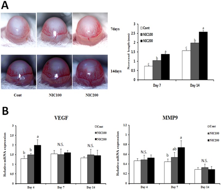
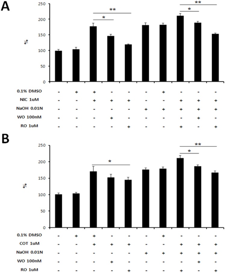
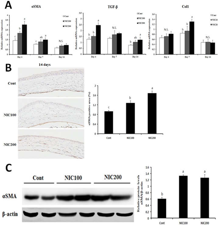
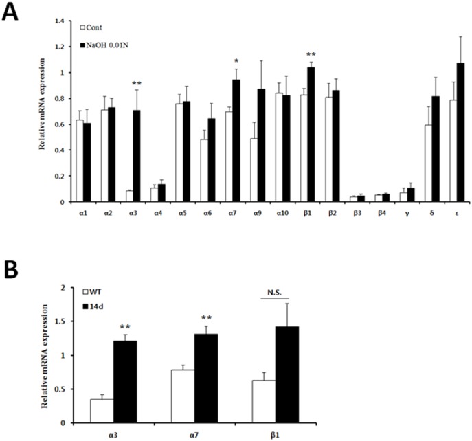
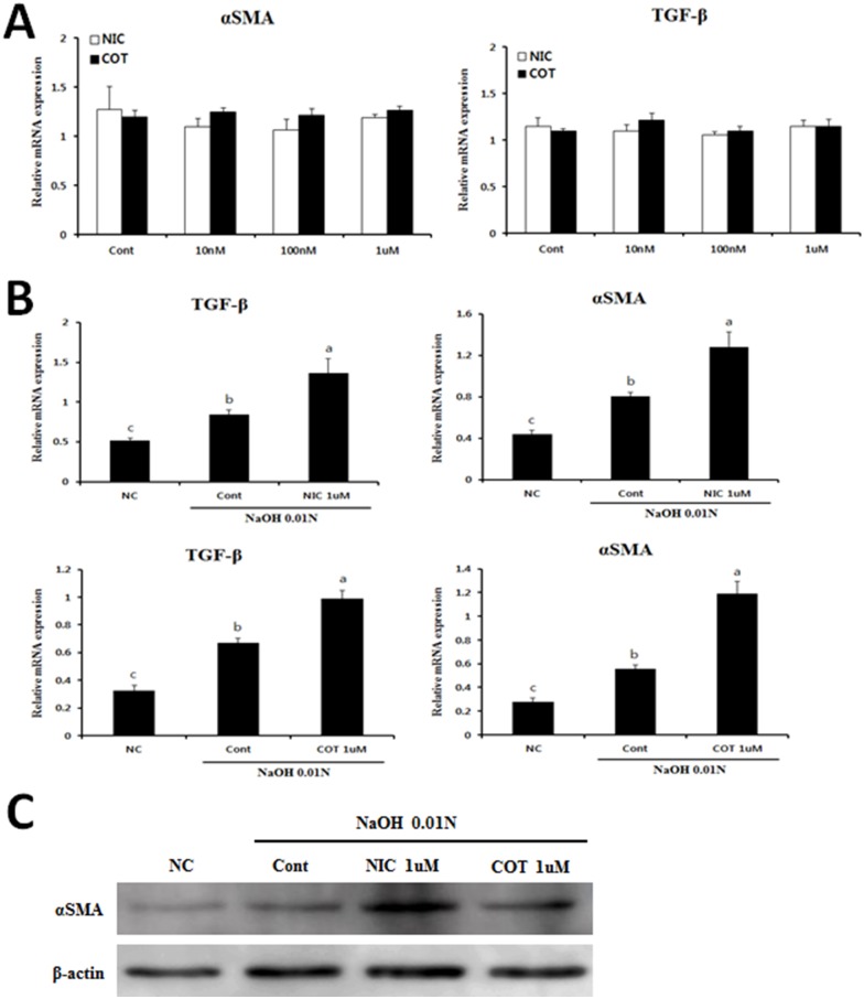
Epidemiological studies have indicated that smoking is a pivotal risk factor for the progression of several chronic diseases. Nicotine, the addictive component of cigarettes, has powerful pathophysiological properties in the body. Although the effects of cigarette smoking on corneal re-epithelialization have been studied, the effects of nicotine on corneal wound healing-related neovascularization and fibrosis have not been fully demonstrated. The aim of this study was to evaluate the effects of chronic administration of nicotine on corneal wound healing following acute insult induced by an alkali burn. BALB/C female mice randomly received either vehicle (2% saccharin) or nicotine (100 or 200 μg/ml in 2% saccharin) in drinking water ad libitum. After 1 week, animals were re-randomized and the experimental group was subjected to a corneal alkali burn, and then nicotine was administered until day 14 after the alkali burn. A corneal alkali burn model was generated by placing a piece of 2 mm-diameter filter paper soaked in 1N NaOH on the right eye. Histopathological analysis and the expression level of the pro-angiogenic genes vascular endothelial growth factor (VEGF) and matrix metalloproteinase-9 (MMP9) revealed that chronic nicotine administration enhanced alkali burn-induced corneal neovascularization. Furthermore, the mRNA expression of the pro-fibrogenic factors α-smooth muscle actin (αSMA), transforming growth factor-β (TGF-β), and collagen α1 (Col1) was enhanced in the high-concentration nicotine-treated group compared with the vehicle group after corneal injury. Immunohistochemical analysis also showed that the αSMA-positive area was increased in chronic nicotine-treated mice after corneal alkali burn. An in vitro assay found that expression of the α3, α7, and β1 nicotinic acetylcholine receptor (nAChR) subunits was significantly increased by chemical injury in human corneal fibroblast cells. Moreover, alkali-induced fibrogenic gene expression and proliferation of fibroblast cells were further increased by treatment with nicotine and cotinine. The proliferation of such cells induced by treatment of nicotine and cotinine was reduced by inhibition of the PI3K and PKC pathways using specific inhibitors. In conclusion, chronic administration of nicotine accelerated the angiogenic and fibrogenic healing processes in alkali-burned corneal tissue.
流行病学研究表明,吸烟是多种慢性疾病进展的关键危险因素。尼古丁作为香烟中的成瘾成分,在体内具有强大的病理生理特性。虽然吸烟对角膜再上皮化的影响已得到研究,但尼古丁对角膜伤口愈合相关的新生血管形成和纤维化的影响尚未完全阐明。本研究的目的是评估长期给予尼古丁对碱烧伤诱导的急性损伤后角膜伤口愈合的影响。BALB/C雌性小鼠随机自由饮用含赋形剂(2%糖精)或尼古丁(2%糖精中含100或200μg/ml)的饮水。1周后,动物重新随机分组,实验组进行角膜碱烧伤,然后在碱烧伤后第14天前持续给予尼古丁。通过将一片浸泡在1N NaOH中的2毫米直径滤纸放置在右眼上建立角膜碱烧伤模型。组织病理学分析以及促血管生成基因血管内皮生长因子(VEGF)和基质金属蛋白酶-9(MMP9)的表达水平显示,长期给予尼古丁可增强碱烧伤诱导的角膜新生血管形成。此外,与赋形剂组相比,高浓度尼古丁处理组在角膜损伤后促纤维化因子α平滑肌肌动蛋白(αSMA)、转化生长因子-β(TGF-β)和胶原蛋白α1(Col1)的mRNA表达增强。免疫组织化学分析还显示,角膜碱烧伤后,长期尼古丁处理的小鼠中αSMA阳性区域增加。体外实验发现,化学损伤可显著增加人角膜成纤维细胞中α3、α7和β1烟碱型乙酰胆碱受体(nAChR)亚基的表达。此外,尼古丁和可替宁处理可进一步增加碱诱导的成纤维细胞纤维化基因表达和增殖。使用特异性抑制剂抑制PI3K和PKC途径可减少尼古丁和可替宁处理诱导的此类细胞增殖。总之,长期给予尼古丁加速了碱烧伤角膜组织的血管生成和纤维化愈合过程。