Jia Yingxian, Shi Xiaohan, Xie Yidong, Xie Xiaochuan, Wang Yan, Li Shangwei
Division of Reproductive Medical Center, West China Second University Hospital, Sichuan University, Chengdu, Sichuan, China.
Key Laboratory of Birth Defects and Related Diseases of Women and Children, West China Second University Hospital of Sichuan University, Chengdu, Sichuan, China.
Stem Cell Res Ther. 2017 Jun 24;8(1):152. doi: 10.1186/s13287-017-0604-4.
To reduce young female fertility loss, the in-vitro culture of cryopreserved ovarian cortical tissues (OCTs) is considered an effective approach without delaying treatment and undergoing stimulation medicine. However, ischemic damage and follicular loss during the in-vitro culture of OCTs are major technical challenges. Human umbilical cord stem cells (HUMSCs) and their conditioned medium (HUMSC-CM) have been considered to be potential resources for regeneration medicine because they secrete cytokines and enhance cell survival and function. The aim of this study was to determine whether HUMSC-CM improves the development of frozen-thawed in-vitro cultured ovarian tissues compared with a serum-free culture medium (SF-CM).
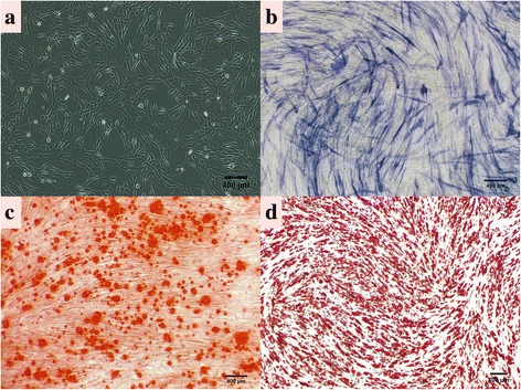
The thawed OCTs (n = 68) were cultivated in HUMSC-CM and SF-CM in vitro for 8 days, and the ovarian tissues were processed and analyzed by a classical histological evaluation. The microvessel density (MVD) and apotosis detection during in-vitro culture of OCTs were also performed.
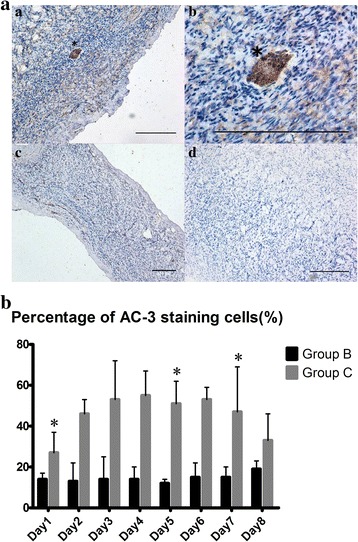
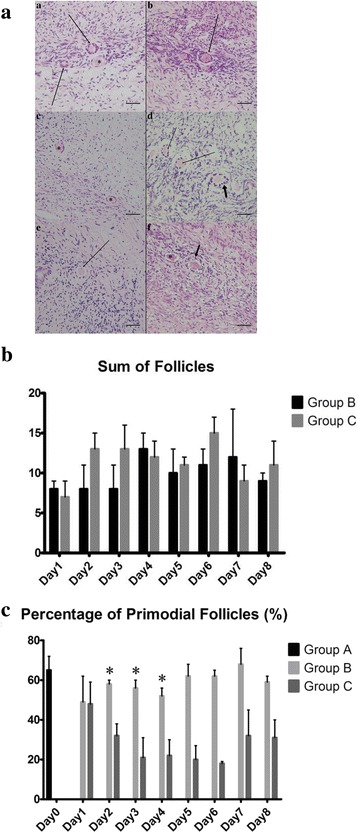
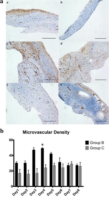
A significant difference in the rate of morphologically normal primordial follicles in the HUMSC-CM group was observed compared to that in the SF-CM group (group C) from days 2 to 4 (day 2: group B 58.0 ± 2.45% vs group C 32.0 ± 5.83%, p = 0.002; day 3: group B 55.5 ± 4.20% vs group C 21.0 ± 9.80%, p = 0.048; day 4: group B 52.0 ± 4.08% vs group C 21.5 ± 8.19%, p = 0.019). The microvessel density (MVD) detection showed a time-dependent increase and peaked on day 4. There was a significant difference between groups B (49.33 ± 0.58) and C (24.33 ± 3.79) (p = 0.036). The percentage of apoptotic follicles in group B was lower than that in group C on day 1 (13.75 ± 2.50% vs 27.0 ± 10.10%, p = 0.003), day 5 (11.75 ± 1.50% vs 51.0 ± 10.5%, p = 0.019) and day 7 (15.0 ± 5.10% vs 46.5 ± 21.75%, p = 0.018).
These data have provided the first experimental evidence of the effect of HUMSC-CM on frozen-thawed OCTs in vitro. The results showed that the HUMSC-CM group provided a better protecting effect on the in-vitro culture of the cryopreserved OCTs compared to the SF-CM group.
为减少年轻女性生育力的损失,冷冻保存的卵巢皮质组织(OCTs)的体外培养被认为是一种有效的方法,无需延迟治疗和使用促排卵药物。然而,OCTs体外培养过程中的缺血损伤和卵泡丢失是主要的技术挑战。人脐带干细胞(HUMSCs)及其条件培养基(HUMSC-CM)被认为是再生医学的潜在资源,因为它们能分泌细胞因子并提高细胞存活率和功能。本研究的目的是确定与无血清培养基(SF-CM)相比,HUMSC-CM是否能改善冻融后体外培养的卵巢组织的发育。
将解冻的OCTs(n = 68)在HUMSC-CM和SF-CM中体外培养8天,然后通过经典组织学评估对卵巢组织进行处理和分析。还对OCTs体外培养过程中的微血管密度(MVD)和凋亡进行了检测。
与SF-CM组(C组)相比,在第2至4天,HUMSC-CM组形态正常的原始卵泡率有显著差异(第2天:B组58.0±2.45% vs C组32.0±5.83%,p = 0.002;第3天:B组55.5±4.20% vs C组21.0±9.80%,p = 0.048;第4天:B组52.0±4.08% vs C组21.5±8.19%,p = 0.019)。微血管密度(MVD)检测显示其随时间增加,并在第4天达到峰值。B组(49.33±0.58)和C组(24.33±3.79)之间存在显著差异(p = 0.036)。第1天(13.75±2.50% vs 27.0±10.10%,p = 0.003)、第5天(11.75±1.50% vs 51.0±10.5%,p = 0.019)和第7天(15.0±5.10% vs 46.5±21.75%,p = 0.018),B组凋亡卵泡的百分比低于C组。
这些数据首次提供了HUMSC-CM对冻融后OCTs体外作用的实验证据。结果表明,与SF-CM组相比,HUMSC-CM组对冷冻保存的OCTs体外培养具有更好的保护作用。