Jakab András, Tuura Ruth, Kellenberger Christian, Scheer Ianina
Center for MR-Research, University Children's Hospital, Zürich, Switzerland; Computational Imaging Research Lab (CIR), Department of Biomedical Imaging and Image-guided Therapy, Medical University of Vienna, Vienna, Austria.
Center for MR-Research, University Children's Hospital, Zürich, Switzerland.
Neuroimage Clin. 2017 Jun 9;15:601-612. doi: 10.1016/j.nicl.2017.06.013. eCollection 2017.
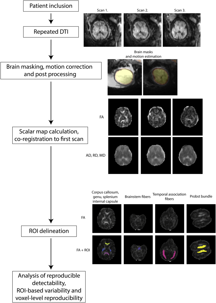
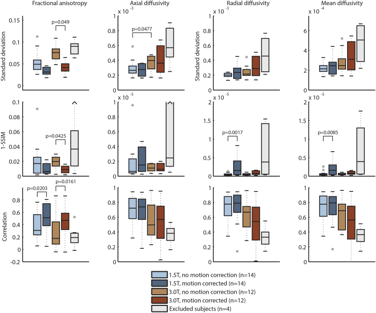
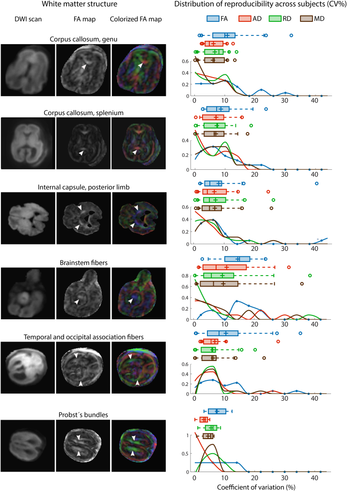
Our purpose was to evaluate the within-subject reproducibility of diffusion tensor imaging (DTI) metrics and the visibility of major white matter structures. Images for 30 fetuses (20-33. postmenstrual weeks, normal neurodevelopment: 6 cases, cerebral pathology: 24 cases) were acquired on 1.5 T or 3.0 T MRI. DTI with 15 diffusion-weighting directions was repeated three times for each case, TR/TE: 2200/63 ms, voxel size: 1 ∗ 1 mm, slice thickness: 3-5 mm, b-factor: 700 s/mm. Reproducibility was evaluated from structure detectability, variability of DTI measures using the coefficient of variation (CV), image correlation and structural similarity across repeated scans for six selected structures. The effect of age, scanner type, presence of pathology was determined using Wilcoxon rank sum test. White matter structures were detectable in the following percentage of fetuses in at least two of the three repeated scans: corpus callosum genu 76%, splenium 64%, internal capsule, posterior limb 60%, brainstem fibers 40% and temporooccipital association pathways 60%. The mean CV of DTI metrics ranged between 3% and 14.6% and we measured higher reproducibility in fetuses with normal brain development. Head motion was negatively correlated with reproducibility, this effect was partially ameliorated by motion-correction algorithm using image registration. Structures on 3.0 T had higher variability both with- and without motion correction. Fetal DTI is reproducible for projection and commissural bundles during mid-gestation, however, in 16-30% of the cases, data were corrupted by artifacts, resulting in impaired detection of white matter structures. To achieve robust results for the quantitative analysis of diffusivity and anisotropy values, fetal-specific image processing is recommended and repeated DTI is needed to ensure the detectability of fiber pathways.
我们的目的是评估扩散张量成像(DTI)指标的受试者内重复性以及主要白质结构的可视性。对30例胎儿(孕龄20 - 33周,正常神经发育:6例,脑病变:24例)进行了1.5T或3.0T磁共振成像(MRI)检查。对每个病例采用15个扩散加权方向进行DTI扫描,重复三次,重复时间/回波时间(TR/TE):2200/63ms,体素大小:1×1mm,层厚:3 - 5mm,b值:700s/mm²。通过结构可检测性、使用变异系数(CV)评估DTI测量值的变异性、重复扫描间的图像相关性以及六个选定结构的结构相似性来评估重复性。使用Wilcoxon秩和检验确定年龄、扫描仪类型、病变存在情况的影响。在三次重复扫描中的至少两次扫描中,以下白质结构在胎儿中的可检测百分比为:胼胝体膝部76%,压部64%,内囊后肢60%,脑干纤维40%,颞枕联合通路60%。DTI指标的平均CV在3%至14.6%之间,我们发现脑发育正常的胎儿具有更高的重复性。头部运动与重复性呈负相关,使用图像配准的运动校正算法可部分改善这种影响。无论有无运动校正,3.0T扫描的结构变异性均更高。孕中期胎儿DTI对投射束和连合束具有可重复性,然而,在16% - 30%的病例中,数据因伪影而受损,导致白质结构检测受损。为了获得关于扩散率和各向异性值定量分析的可靠结果,建议采用胎儿特异性图像处理方法,并需要重复进行DTI检查以确保纤维束的可检测性。