Attia Amalina Binte Ebrahim, Chuah Sai Yee, Razansky Daniel, Ho Chris Jun Hui, Malempati Pinky, Dinish U S, Bi Renzhe, Fu Chit Yaw, Ford Steven J, Lee Joyce Siong-See, Tan Melissa Wee Ping, Olivo Malini, Thng Steven Tien Guan
Bio-optical Imaging Group, Singapore Bioimaging Consortium, ASTAR, Singapore.
National Skin Centre, Singapore.
Photoacoustics. 2017 Jun 4;7:20-26. doi: 10.1016/j.pacs.2017.05.003. eCollection 2017 Sep.
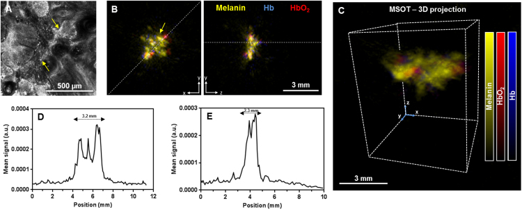
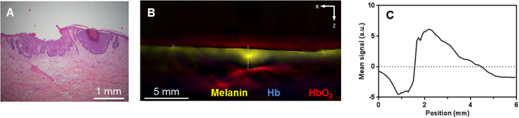
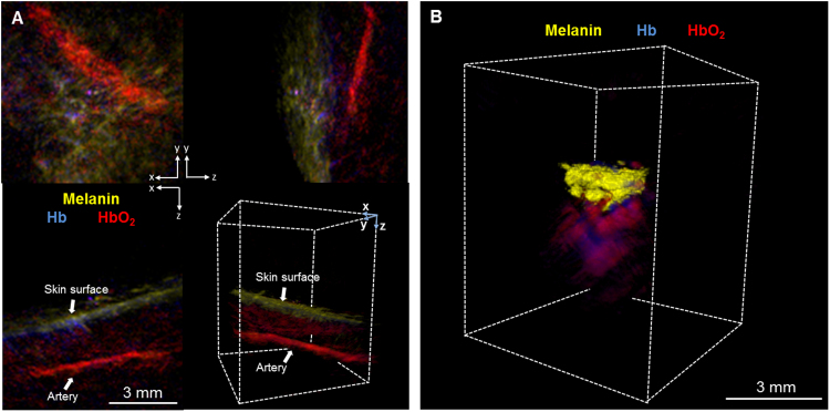
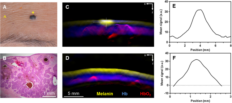
Currently, imaging technologies that enable dermsurgeons to visualize non-melanoma skin cancers (NMSC) preoperatively are lacking, resulting in excessive or incomplete removal. Multispectral optoacoustic tomography (MSOT) is a volumetric imaging tool to differentiate tissue chromophores and exogenous contrast agents, based on differences in their spectral signatures and used for high-resolution imaging of functional and molecular contrast at centimeter scale depth. We performed MSOT imaging with two- and three-dimensional handheld scanners on 21 Asian patients with NMSC. The tumors and their oxygenation parameters could be distinguished from normal skin endogenously. The lesion dimensions and depths were extracted from the spectral melanin component with three-dimensional spatial resolution up to 80 μm. The intraclass correlation coefficient correlating tumor dimension measurements between MSOT and histology of excised tumors, showed good correlation. Real-time 3D imaging was found to provide information on lesion morphology and its underlying neovasculature, indicators of the tumor's aggressiveness.
目前,能够让皮肤科医生在术前可视化非黑色素瘤皮肤癌(NMSC)的成像技术尚不完善,这导致切除过度或不彻底。多光谱光声断层扫描(MSOT)是一种容积成像工具,可根据组织发色团和外源性造影剂的光谱特征差异来区分它们,并用于在厘米级深度进行功能和分子对比的高分辨率成像。我们使用二维和三维手持式扫描仪对21例患有NMSC的亚洲患者进行了MSOT成像。肿瘤及其氧合参数可以从正常皮肤中内源性地区分出来。通过三维空间分辨率高达80μm的光谱黑色素成分提取病变的尺寸和深度。MSOT测量的肿瘤尺寸与切除肿瘤的组织学之间的组内相关系数显示出良好的相关性。发现实时三维成像可提供有关病变形态及其潜在新生血管的信息,这些都是肿瘤侵袭性的指标。