Townes-Anderson Ellen, Wang Jianfeng, Halász Éva, Sugino Ilene, Pitler Amy, Whitehead Ian, Zarbin Marco
Department of Pharmacology, Physiology, and Neuroscience, Rutgers New Jersey Medical School, Newark, NJ, USA.
Institute of Ophthalmology and Visual Science, Rutgers New Jersey Medical School, Newark, NJ, USA.
Transl Vis Sci Technol. 2017 Jun 20;6(3):22. doi: 10.1167/tvst.6.3.22. eCollection 2017 Jun.
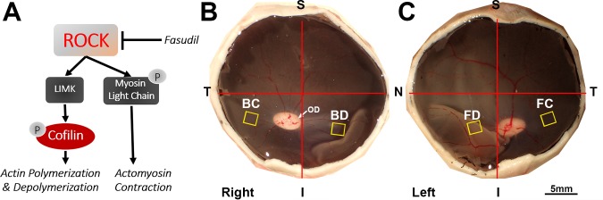
Retinal detachment disrupts the rod-bipolar synapse in the outer plexiform layer by retraction of rod axons. We showed that breakage is due to RhoA activation whereas inhibition of Rho kinase (ROCK), using Y27632, reduces synaptic damage. We test whether the ROCK inhibitor fasudil, used for other clinical applications, can prevent synaptic injury after detachment.
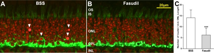
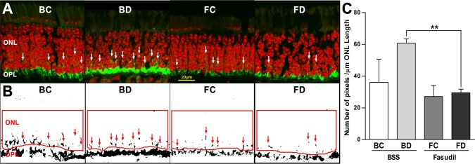
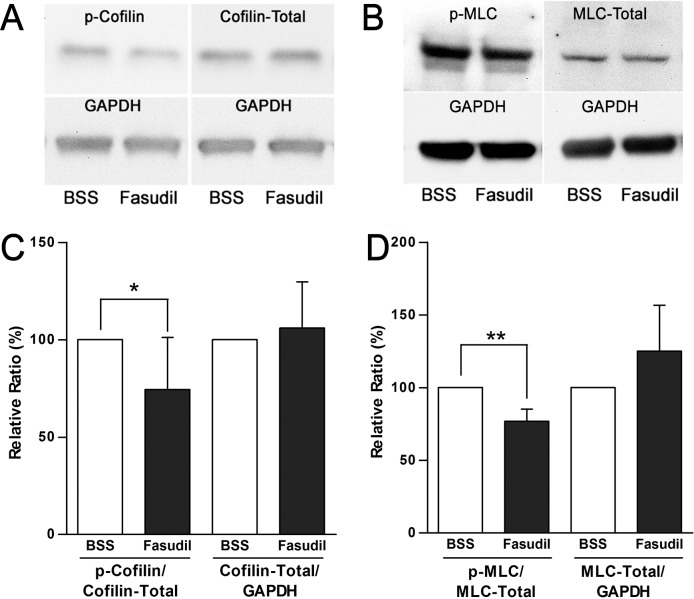
Detachments were made in pigs by subretinal injection of balanced salt solution (BSS) or fasudil (1, 10 mM). In some animals, fasudil was injected intravitreally after BSS-induced detachment. After 2 to 4 hours, retinae were fixed for immunocytochemistry and confocal microscopy. Axon retraction was quantified by imaging synaptic vesicle label in the outer nuclear layer. Apoptosis was analyzed using propidium iodide staining. For biochemical analysis by Western blotting, retinal explants, detached from retinal pigmented epithelium, were cultured for 2 hours.
Subretinal injection of fasudil (10 mM) reduced retraction of rod spherules by 51.3% compared to control detachments ( = 3 pigs, = 0.002). Intravitreal injection of 10 mM fasudil, a more clinically feasible route of administration, also reduced retraction (28.7%, = 5, < 0.05). Controls had no photoreceptor degeneration at 2 hours, but by 4 hours apoptosis was evident. Fasudil 10 mM reduced pyknotic nuclei by 55.7% ( = 4, < 0.001). Phosphorylation of cofilin and myosin light chain, downstream effectors of ROCK, was decreased with 30 μM fasudil ( = 8-10 explants, < 0.05).
Inhibition of ROCK signaling with fasudil reduced photoreceptor degeneration and preserved the rod-bipolar synapse after retinal detachment.
These results support the possibility, previously tested with Y27632, that ROCK inhibition may attenuate synaptic damage in iatrogenic detachments.
视网膜脱离通过视杆轴突回缩破坏外网状层中的视杆 - 双极突触。我们发现这种破坏是由于RhoA激活所致,而使用Y27632抑制Rho激酶(ROCK)可减少突触损伤。我们测试用于其他临床应用的ROCK抑制剂法舒地尔是否能预防视网膜脱离后的突触损伤。
通过视网膜下注射平衡盐溶液(BSS)或法舒地尔(1、10 mM)在猪中造成视网膜脱离。在一些动物中,在BSS诱导的视网膜脱离后玻璃体内注射法舒地尔。2至4小时后,固定视网膜用于免疫细胞化学和共聚焦显微镜检查。通过对外核层中突触小泡标记成像来量化轴突回缩。使用碘化丙啶染色分析细胞凋亡。对于通过蛋白质印迹进行的生化分析,将从视网膜色素上皮分离的视网膜外植体培养2小时。
与对照性视网膜脱离相比,视网膜下注射法舒地尔(10 mM)使视杆小球回缩减少了51.3%(n = 3头猪,P = 0.002)。玻璃体内注射10 mM法舒地尔,这是一种临床上更可行的给药途径,也减少了回缩(28.7%,n = 5,P < 0.05)。对照组在2小时时没有光感受器变性,但到4小时时细胞凋亡明显。10 mM法舒地尔使固缩核减少了55.7%(n = 4,P < 0.001)。ROCK的下游效应分子cofilin和肌球蛋白轻链的磷酸化在30 μM法舒地尔作用下降低(n = 8 - 10个外植体,P < 0.05)。
用法舒地尔抑制ROCK信号传导可减少视网膜脱离后的光感受器变性并保留视杆 - 双极突触。
这些结果支持了之前用Y27632测试过的可能性,即ROCK抑制可能减轻医源性视网膜脱离中的突触损伤。