Takeda Tsutomu, Asaoka Daisuke, Tajima Yuzuru, Matsumoto Kenshi, Takeda Naoto, Hiromoto Takahumi, Okubo Shoki, Saito Hiroaki, Aoyama Tomonori, Shibuya Tomoyoshi, Sakamoto Naoto, Hojo Mariko, Osada Taro, Nagahara Akihito, Yao Takashi, Watanabe Sumio
Department of Gastroenterology, Juntendo University School of Medicine, Tokyo, Japan.
Department of Human Pathology, Juntendo University School of Medicine, Tokyo, Japan.
Clin J Gastroenterol. 2017 Oct;10(5):478-484. doi: 10.1007/s12328-017-0756-x. Epub 2017 Jun 28.
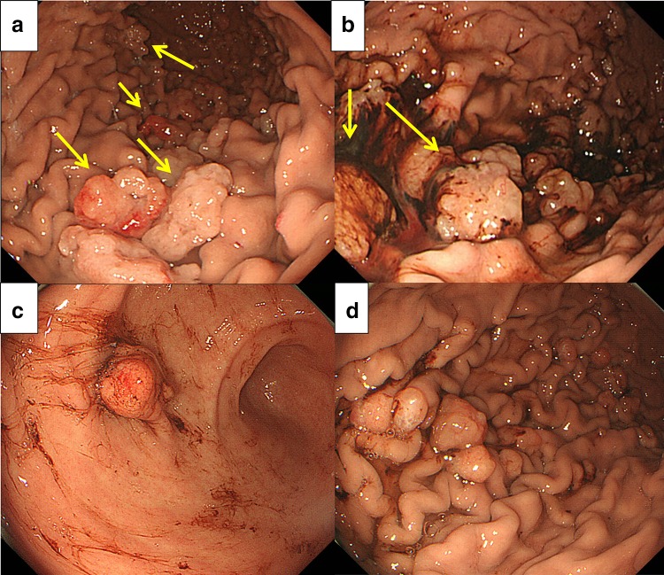
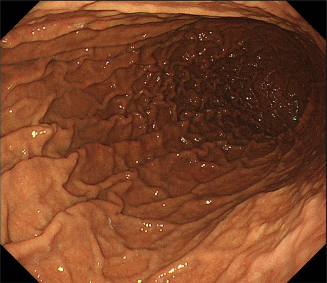
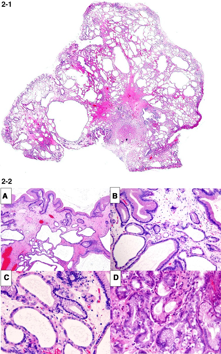
We report a rare case of hemorrhagic gastric polyps resulting in anemia during long-term proton pump inhibitor (PPI) administration that endoscopically looked like a fundic gland polyp (FGP). A 44-year-old man presented complaining of anemia and tarry stools. Esophagogastroduodenoscopy (EGD) demonstrated multiple white edematous polyps in the corpus and antrum, which were considered to be FGPs. We attempted endoscopic hemostasis but hemorrhaging increased because of hemorrhagic polyps and vulnerable gastric mucosa. Re-bleeding occurred several times. Polyp resection was performed at 24 polyp sites. We also ceased the administration of PPI. Microscopically, polyps showed characteristics of hyperplasia in the foveolar epithelium, extensions of fundic glands, and edema of the stroma. The proliferation of parietal and chief cells was also observed. Immunohistochemically, aquaporin-4 (AQP4) and KCNQ1-positive parietal cells and dilated mucous glands were found from the basal side to the apical side of the mucosa. These findings were compatible with the development of lesions associated with the long-term administration of PPI. EGD revealed an improvement in the vulnerability of gastric mucosa and the development of polyps, with no further gastric polyps observed 1 year after discharge. Bleeding from polyps resembling FGPs is generally rare, with indications that long-term PPI administration may induce such bleeding.
我们报告了一例罕见的出血性胃息肉病例,该病例在长期服用质子泵抑制剂(PPI)期间导致贫血,内镜检查时其外观类似胃底腺息肉(FGP)。一名44岁男性因贫血和柏油样便前来就诊。食管胃十二指肠镜检查(EGD)显示胃体和胃窦有多个白色水肿性息肉,被认为是胃底腺息肉。我们尝试进行内镜止血,但由于出血性息肉和脆弱的胃黏膜,出血反而增加。再次出血发生了几次。在24个息肉部位进行了息肉切除术。我们还停止了PPI的给药。显微镜下,息肉表现为小凹上皮增生、胃底腺延伸和间质水肿的特征。还观察到壁细胞和主细胞的增殖。免疫组织化学检查发现,从黏膜基侧到顶端,水通道蛋白-4(AQP4)和KCNQ1阳性的壁细胞以及扩张的黏液腺均有发现。这些发现与长期服用PPI相关病变的发展相符。EGD显示胃黏膜的脆弱性有所改善,息肉也有发展,出院1年后未再观察到胃息肉。类似胃底腺息肉的息肉出血通常很少见,有迹象表明长期服用PPI可能会引发此类出血。