Zitzelsberger Tanja, Eigentler Thomas K, Krumm Patrick, Nikolaou Konstantin, Garbe Claus, Gawaz Meinrad, Klumpp Bernhard
Department of Diagnostic and Interventional Radiology, Eberhard Karls University Tuebingen, Hoppe-Seyler-Straße 3, 72076, Tuebingen, Germany.
Eberhard-Karls-University Tuebingen, Center for Dermatooncology, Liebermeisterstr. 25, 72076, Tuebingen, Germany.
Cancer Imaging. 2017 Jul 1;17(1):19. doi: 10.1186/s40644-017-0122-8.
Due to prolonged survival and technical advances in CT imaging, cardiac metastases in patients with malignant melanoma are observed more frequently nowadays. The aim of the present study was to assess the anatomic distribution as well as the morphologic and histologic appearance of cardiac metastases from malignant melanoma.
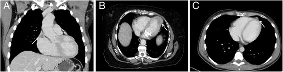
Twenty five patients with known metastasized melanoma and with incidental finding of cardiac metastases during routine staging CT were retrospectively included in this study. CT images were assessed for the presence, localization and extent of cardiac metastases. Histological results, mutational analysis and tumor markers were reviewed.
Fourteen out of 25 patients presented with singular cardiac mass (56%), whereas ten patients (40%) presented with multifocal and one patient with disseminated cardiac metastases. Twelve patients presented with endocardial (48%), eight with myocardial and two with pericardial metastases. Most frequent site involved in endocardial metastases was the right atrium (67%) followed by the right ventricle (33%). There seems to be a correlation between histological subtype and location of cardiac metastasis. Median survival after diagnosis of cardiac metastases was 8 months, with no significant difference regarding the localization of metastases within the heart.
Cardiac metastases can involve every part of the heart possibly in dependence of histological subtype. The awareness of different types of cardiac metastases and their characteristic appearance on CT images is necessary for further investigations and might contribute to targeted therapy.
由于生存期延长以及CT成像技术的进步,如今恶性黑色素瘤患者的心脏转移更为常见。本研究的目的是评估恶性黑色素瘤心脏转移的解剖分布以及形态学和组织学表现。
本研究回顾性纳入了25例已知有转移性黑色素瘤且在常规分期CT检查中偶然发现心脏转移的患者。对CT图像进行评估,以确定心脏转移的存在、定位和范围。回顾了组织学结果、突变分析和肿瘤标志物。
25例患者中有14例出现单发心脏肿块(56%),10例(40%)出现多灶性心脏转移,1例出现弥漫性心脏转移。12例患者出现心内膜转移(48%),8例出现心肌转移,2例出现心包转移。心内膜转移最常累及的部位是右心房(67%),其次是右心室(33%)。心脏转移的组织学亚型与位置之间似乎存在相关性。心脏转移诊断后的中位生存期为8个月,心脏内转移部位之间无显著差异。
心脏转移可能累及心脏的各个部位,这可能取决于组织学亚型。了解不同类型的心脏转移及其在CT图像上的特征表现对于进一步研究很有必要,并且可能有助于靶向治疗。