• 文献检索
  • 文档翻译
  • 深度研究
  • 学术资讯
  • Suppr Zotero 插件Zotero 插件
  • 邀请有礼
  • 套餐&价格
  • 历史记录
应用&插件
Suppr Zotero 插件Zotero 插件浏览器插件Mac 客户端Windows 客户端微信小程序
定价
高级版会员购买积分包购买API积分包
服务
文献检索文档翻译深度研究API 文档MCP 服务
关于我们
关于 Suppr公司介绍联系我们用户协议隐私条款
关注我们

Suppr 超能文献

核心技术专利:CN118964589B侵权必究
粤ICP备2023148730 号-1Suppr @ 2026

文献检索

告别复杂PubMed语法,用中文像聊天一样搜索,搜遍4000万医学文献。AI智能推荐,让科研检索更轻松。

立即免费搜索

文件翻译

保留排版,准确专业,支持PDF/Word/PPT等文件格式,支持 12+语言互译。

免费翻译文档

深度研究

AI帮你快速写综述,25分钟生成高质量综述,智能提取关键信息,辅助科研写作。

立即免费体验

胼胝体压部病变:疾病谱与MRI表现

Splenial Lesions of the Corpus Callosum: Disease Spectrum and MRI Findings.

作者信息

Park Sung Eun, Choi Dae Seob, Shin Hwa Seon, Baek Hye Jin, Choi Ho Cheol, Kim Ji Eun, Choi Hye Young, Park Mi Jung

机构信息

Department of Radiology, Gyeongsang National University School of Medicine, Jinju 52727, Korea.

Gyeongsang Institute of Health Science, Gyeongsang National University School of Medicine, Jinju 52727, Korea.

出版信息

Korean J Radiol. 2017 Jul-Aug;18(4):710-721. doi: 10.3348/kjr.2017.18.4.710. Epub 2017 May 19.

DOI:10.3348/kjr.2017.18.4.710
PMID:28670166
原文链接:https://pmc.ncbi.nlm.nih.gov/articles/PMC5447647/
Abstract

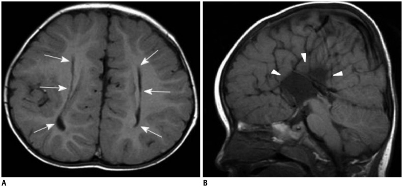
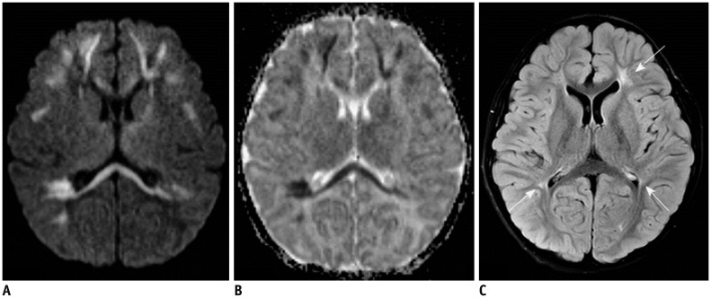
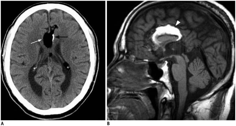
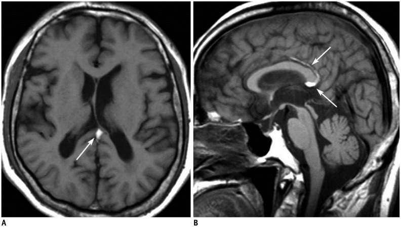
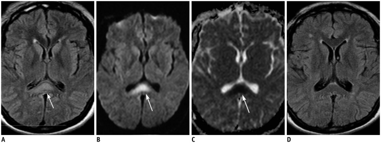
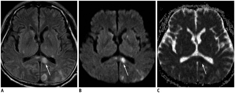
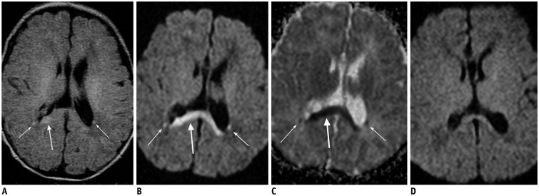
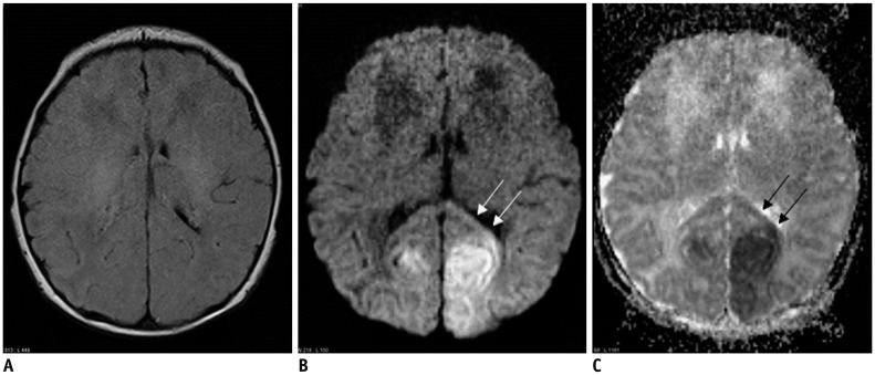
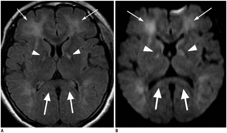
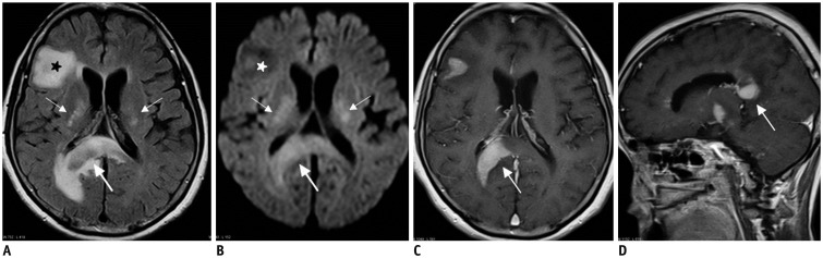
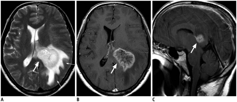
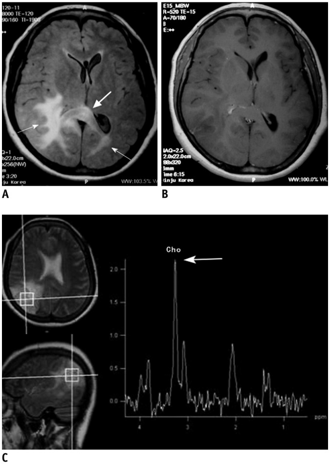
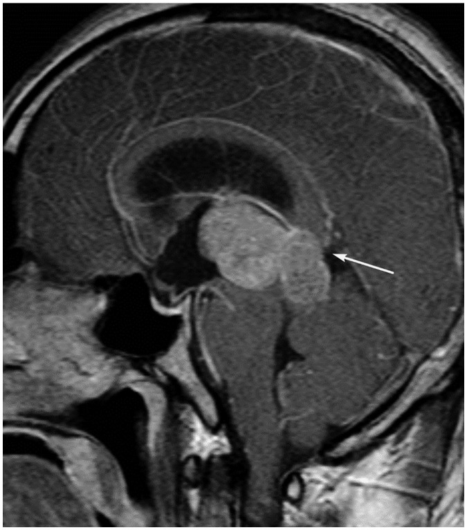
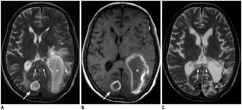
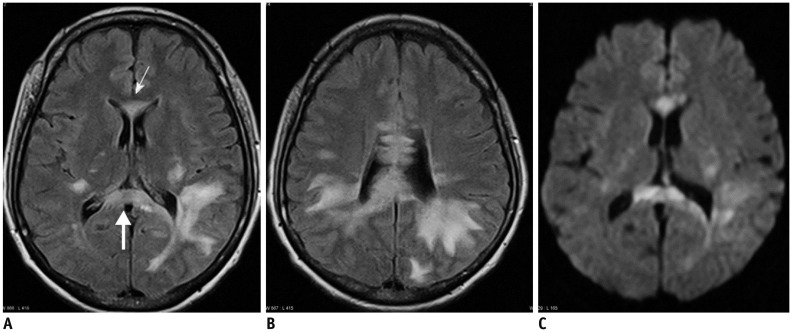
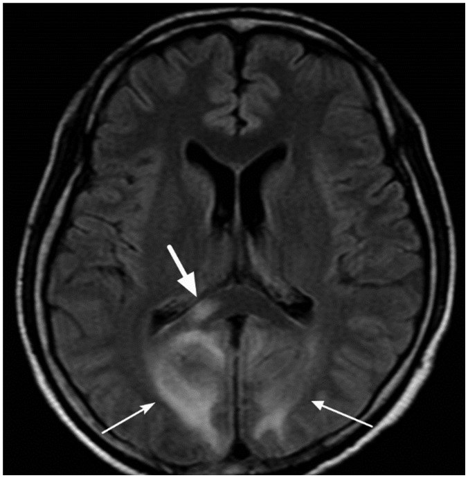
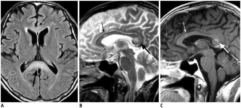
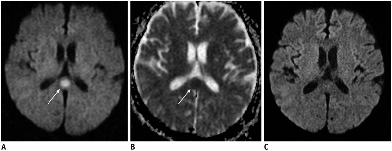
The corpus callosum (CC) is the largest white matter structure in the brain, consisting of more than 200-250 million axons that provide a large connection mainly between homologous cerebral cortical areas in mirror image sites. The posterior end of the CC is the thickest part, which is called the splenium. Various diseases including congenital to acquired lesions including congenital anomalies, traumatic lesions, ischemic diseases, tumors, metabolic, toxic, degenerative, and demyelinating diseases, can involve the splenium of the CC and their clinical symptoms and signs are also variable. Therefore, knowledge of the disease entities and the imaging findings of lesions involving the splenium is valuable in clinical practice. MR imaging is useful for the detection and differential diagnosis of splenial lesions of the CC. In this study, we classify the disease entities and describe imaging findings of lesions involving the splenium of the CC based on our experiences and a review of the literature.

摘要

胼胝体(CC)是大脑中最大的白质结构,由超过2亿到2.5亿条轴突组成,主要在镜像部位的同源大脑皮质区域之间提供大量连接。CC的后端是最厚的部分,称为压部。包括先天性到后天性病变,如先天性异常、创伤性病变、缺血性疾病、肿瘤、代谢性、中毒性、退行性和脱髓鞘疾病等各种疾病,都可能累及CC的压部,其临床症状和体征也各不相同。因此,了解累及压部的疾病实体和病变的影像学表现在临床实践中具有重要价值。磁共振成像(MR)对CC压部病变的检测和鉴别诊断很有用。在本研究中,我们根据经验和文献回顾,对疾病实体进行分类,并描述累及CC压部病变的影像学表现。

https://cdn.ncbi.nlm.nih.gov/pmc/blobs/cd19/5447647/e6dbe22aa240/kjr-18-710-g018.jpg
https://cdn.ncbi.nlm.nih.gov/pmc/blobs/cd19/5447647/5f4e899e0c49/kjr-18-710-g001.jpg
https://cdn.ncbi.nlm.nih.gov/pmc/blobs/cd19/5447647/8fc54a8b7d42/kjr-18-710-g002.jpg
https://cdn.ncbi.nlm.nih.gov/pmc/blobs/cd19/5447647/ff5acc316140/kjr-18-710-g003.jpg
https://cdn.ncbi.nlm.nih.gov/pmc/blobs/cd19/5447647/cde18495f1ec/kjr-18-710-g004.jpg
https://cdn.ncbi.nlm.nih.gov/pmc/blobs/cd19/5447647/e110f645549c/kjr-18-710-g005.jpg
https://cdn.ncbi.nlm.nih.gov/pmc/blobs/cd19/5447647/7e6079d290af/kjr-18-710-g006.jpg
https://cdn.ncbi.nlm.nih.gov/pmc/blobs/cd19/5447647/9562652aa753/kjr-18-710-g007.jpg
https://cdn.ncbi.nlm.nih.gov/pmc/blobs/cd19/5447647/b1c6071a4f3b/kjr-18-710-g008.jpg
https://cdn.ncbi.nlm.nih.gov/pmc/blobs/cd19/5447647/12758249076f/kjr-18-710-g009.jpg
https://cdn.ncbi.nlm.nih.gov/pmc/blobs/cd19/5447647/60730076d8e4/kjr-18-710-g010.jpg
https://cdn.ncbi.nlm.nih.gov/pmc/blobs/cd19/5447647/7705e4033513/kjr-18-710-g011.jpg
https://cdn.ncbi.nlm.nih.gov/pmc/blobs/cd19/5447647/2bca58c722d2/kjr-18-710-g012.jpg
https://cdn.ncbi.nlm.nih.gov/pmc/blobs/cd19/5447647/f07e3a62327b/kjr-18-710-g013.jpg
https://cdn.ncbi.nlm.nih.gov/pmc/blobs/cd19/5447647/4db8dd751716/kjr-18-710-g014.jpg
https://cdn.ncbi.nlm.nih.gov/pmc/blobs/cd19/5447647/0f4fedad0dea/kjr-18-710-g015.jpg
https://cdn.ncbi.nlm.nih.gov/pmc/blobs/cd19/5447647/2204436e445a/kjr-18-710-g016.jpg
https://cdn.ncbi.nlm.nih.gov/pmc/blobs/cd19/5447647/7debe6eb841d/kjr-18-710-g017.jpg
https://cdn.ncbi.nlm.nih.gov/pmc/blobs/cd19/5447647/e6dbe22aa240/kjr-18-710-g018.jpg

相似文献

1
Splenial Lesions of the Corpus Callosum: Disease Spectrum and MRI Findings.胼胝体压部病变:疾病谱与MRI表现
Korean J Radiol. 2017 Jul-Aug;18(4):710-721. doi: 10.3348/kjr.2017.18.4.710. Epub 2017 May 19.
2
[Value of Magnetic Resonance Imaging Findings in Differential Diagnosis between the Adult Reversible Splenial Lesion Syndrome and Ischemic Infarction of the Splenium of the Corpus Callosum].[磁共振成像表现对成人可逆性胼胝体压部病变综合征与胼胝体压部缺血性梗死鉴别诊断的价值]
Zhongguo Yi Xue Ke Xue Yuan Xue Bao. 2018 Apr 28;40(2):233-241. doi: 10.3881/j.issn.1000-503X.2018.02.014.
3
Lesions in the Splenium of the Corpus Callosum on MRI in Children: A Review.儿童胼胝体压部MRI病变综述
J Neuroimaging. 2017 Nov;27(6):549-561. doi: 10.1111/jon.12455. Epub 2017 Jun 27.
4
The splenium of the corpus callosum: embryology, anatomy, function and imaging with pathophysiological hypothesis.胼胝体压部:胚胎学、解剖学、功能与影像学表现及病理生理学假说。
Neuroradiology. 2020 May;62(5):563-585. doi: 10.1007/s00234-019-02357-z. Epub 2020 Feb 15.
5
Transient Splenial Lesion of the Corpus Callosum Related to Migraine with Aura in a Pediatric Patient.一名儿科患者中与伴先兆偏头痛相关的胼胝体短暂性脾周病变
Acta Medica (Hradec Kralove). 2016;59(2):64-6. doi: 10.14712/18059694.2016.91.
6
[Imaging pathologies of the corpus callosum].[胼胝体的影像学病变]
Nervenarzt. 2022 Aug;93(8):844-853. doi: 10.1007/s00115-022-01347-6. Epub 2022 Jul 13.
7
High-resolution imaging of distinct human corpus callosum microstructure and topography of structural connectivity to cortices at high field.高分辨率成像技术可清晰显示人类胼胝体的微观结构和连接皮质的结构连接的拓扑结构,在高场强下进行。
Brain Struct Funct. 2019 Mar;224(2):949-960. doi: 10.1007/s00429-018-1804-0. Epub 2018 Dec 3.
8
Transient Splenial Lesion of the Corpus Callosum After Cabergoline Treatment.卡麦角林治疗后胼胝体一过性孤立性病变
World Neurosurg. 2018 Jun;114:257-258. doi: 10.1016/j.wneu.2018.03.166. Epub 2018 Mar 31.
9
Reversible splenial lesions during febrile illness with or without white matter lesions.发热伴或不伴白质病变时可逆性胼胝体病变。
Brain Dev. 2021 Jun;43(6):698-704. doi: 10.1016/j.braindev.2021.02.004. Epub 2021 Mar 11.
10
Cytotoxic lesions of the corpus callosum in children: Etiology, clinical and radiological features, and prognosis.儿童胼胝体细胞毒性病变:病因、临床和影像学特征及预后。
Brain Dev. 2021 Oct;43(9):919-930. doi: 10.1016/j.braindev.2021.05.001. Epub 2021 Jun 11.

引用本文的文献

1
Clinical analysis of Marchiafava-Bignami disease.Marchiafava-Bignami 病的临床分析。
BMC Neurol. 2024 Oct 14;24(1):389. doi: 10.1186/s12883-024-03901-y.
2
Case report: Symmetrical and increased lateral sway-based walking training for patients with corpus callosum infarction: a case series.病例报告:胼胝体梗死患者基于侧向摆动对称增加的步行训练:病例系列
Front Neurol. 2024 Jun 24;15:1330975. doi: 10.3389/fneur.2024.1330975. eCollection 2024.
3
Neuroimaging features of psilocybin-induced toxic-metabolic encephalopathy in an adolescent.

本文引用的文献

1
Emerging Techniques in Brain Tumor Imaging: What Radiologists Need to Know.脑肿瘤成像的新兴技术:放射科医生需要了解的内容。
Korean J Radiol. 2016 Sep-Oct;17(5):598-619. doi: 10.3348/kjr.2016.17.5.598. Epub 2016 Aug 23.
2
Neonatal parechovirus leucoencephalitis- radiological pattern mimicking hypoxic-ischemic encephalopathy.新生儿细小病毒白质脑病——影像学表现酷似缺氧缺血性脑病
Eur J Radiol. 2016 Feb;85(2):428-34. doi: 10.1016/j.ejrad.2015.11.038. Epub 2015 Dec 2.
3
Infarction of the corpus callosum: a retrospective clinical investigation.
一名青少年中裸盖菇素所致中毒代谢性脑病的神经影像学特征
BMJ Case Rep. 2024 Mar 5;17(3):e259721. doi: 10.1136/bcr-2024-259721.
4
Case report: Susac syndrome-two ends of the spectrum, single center case reports and review of the literature.病例报告:Susac综合征——疾病谱的两端,单中心病例报告及文献综述
Front Neurol. 2024 Feb 16;15:1339438. doi: 10.3389/fneur.2024.1339438. eCollection 2024.
5
Diffusion Restriction in the Splenium: A Comparative Study of Cytotoxic Lesions of the Corpus Callosum (CLOCCs) versus Lesions of Vascular Etiology.胼胝体压部的扩散受限:胼胝体细胞毒性病变(CLOCCs)与血管源性病变的对比研究
J Clin Med. 2023 Nov 8;12(22):6979. doi: 10.3390/jcm12226979.
6
Diffusion-Restricted Lesions of the Splenium: Clinical Presentation, Radiographic Patterns, and Patient Outcomes.胼胝体压部的扩散受限性病变:临床表现、影像学特征及患者预后
Neurol Clin Pract. 2023 Oct;13(5):e200196. doi: 10.1212/CPJ.0000000000200196. Epub 2023 Sep 8.
7
A case of intravascular lymphoma presenting with a lesion in the splenium of the corpus callosum.1例以胼胝体压部病变为表现的血管内淋巴瘤。
Radiol Case Rep. 2023 Mar 16;18(5):1929-1932. doi: 10.1016/j.radcr.2023.02.025. eCollection 2023 May.
8
Clinical Reasoning: A 47-Year-Old Man With an Upper Respiratory Infection, Acute Confusion, Dysarthria, and Ataxia.临床推理:一名 47 岁男性,上呼吸道感染,急性意识混乱,构音障碍和共济失调。
Neurology. 2023 May 16;100(20):978-983. doi: 10.1212/WNL.0000000000206864. Epub 2023 Jan 25.
9
[Imaging pathologies of the corpus callosum].[胼胝体的影像学病变]
Radiologie (Heidelb). 2023 Mar;63(3):209-219. doi: 10.1007/s00117-022-01111-6.
10
Imaging findings of two patients with isolated infarction of the splenium during COVID-19.两例新冠肺炎患者出现单纯胼胝体梗死的影像学表现。
SAGE Open Med Case Rep. 2022 Oct 31;10:2050313X221135232. doi: 10.1177/2050313X221135232. eCollection 2022.
胼胝体梗死:一项回顾性临床研究。
PLoS One. 2015 Mar 18;10(3):e0120409. doi: 10.1371/journal.pone.0120409. eCollection 2015.
4
Distinctive pattern of white matter injury in neonates with rotavirus infection.轮状病毒感染新生儿的白质损伤的特征性模式。
Neurology. 2015 Jan 6;84(1):21-7. doi: 10.1212/WNL.0000000000001107. Epub 2014 Dec 3.
5
Transient restricted diffusion in the splenium of the corpus callosum after brain surgery.脑手术后胼胝体压部的短暂性扩散受限
JBR-BTR. 2013 Mar-Apr;96(2):92. doi: 10.5334/jbr-btr.221.
6
The role of MR imaging in assessment of brain damage from carbon monoxide poisoning: a review of the literature.磁共振成像在评估一氧化碳中毒性脑损伤中的作用:文献综述。
AJNR Am J Neuroradiol. 2014 Apr;35(4):625-31. doi: 10.3174/ajnr.A3489. Epub 2013 Apr 18.
7
Lesions of the corpus callosum.胼胝体病变。
AJR Am J Roentgenol. 2013 Jan;200(1):W1-16. doi: 10.2214/AJR.11.8080.
8
Boomerang sign: Clinical significance of transient lesion in splenium of corpus callosum.回旋镖征:胼胝体压部短暂性病变的临床意义。
Ann Indian Acad Neurol. 2012 Apr;15(2):151-7. doi: 10.4103/0972-2327.95005.
9
Intracranial lesions with high signal intensity on T1-weighted MR images: differential diagnosis.颅内 T1 加权 MR 图像高信号病灶:鉴别诊断。
Radiographics. 2012 Mar-Apr;32(2):499-516. doi: 10.1148/rg.322105761.
10
Imaging of traumatic brain injury: a review of the recent medical literature.创伤性脑损伤的影像学:对近期医学文献的综述。
AJR Am J Roentgenol. 2010 Jan;194(1):16-9. doi: 10.2214/AJR.09.3687.