Mózes Miklós M, Szoleczky Petra, Rosivall László, Kökény Gábor
Institute of Pathophysiology, Semmelweis University, Nagyvárad tér 4, Budapest, H-1089, Hungary.
Hungarian Academy of Sciences and Semmelweis University Research Group for Pediatrics and Nephrology, Budapest, Hungary.
BMC Nephrol. 2017 Jul 3;18(1):209. doi: 10.1186/s12882-017-0626-2.
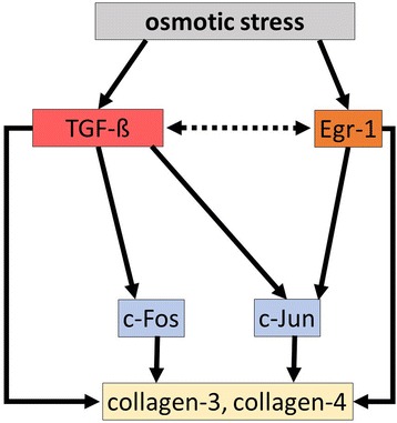
Although TGF-ß and the transcription factor Egr-1 play an important role in both kidney fibrosis and in response to acute changes of renal medullary osmolarity, their role under sustained hypo- or hyperosmolar conditions has not been elucidated. We investigated the effects of chronic hypertonicity and hypotonicity on the renal medullary TGF-ß and Egr-1 expression.
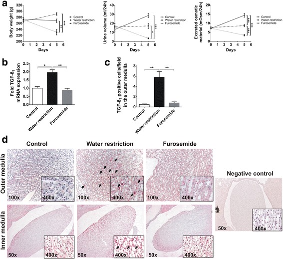
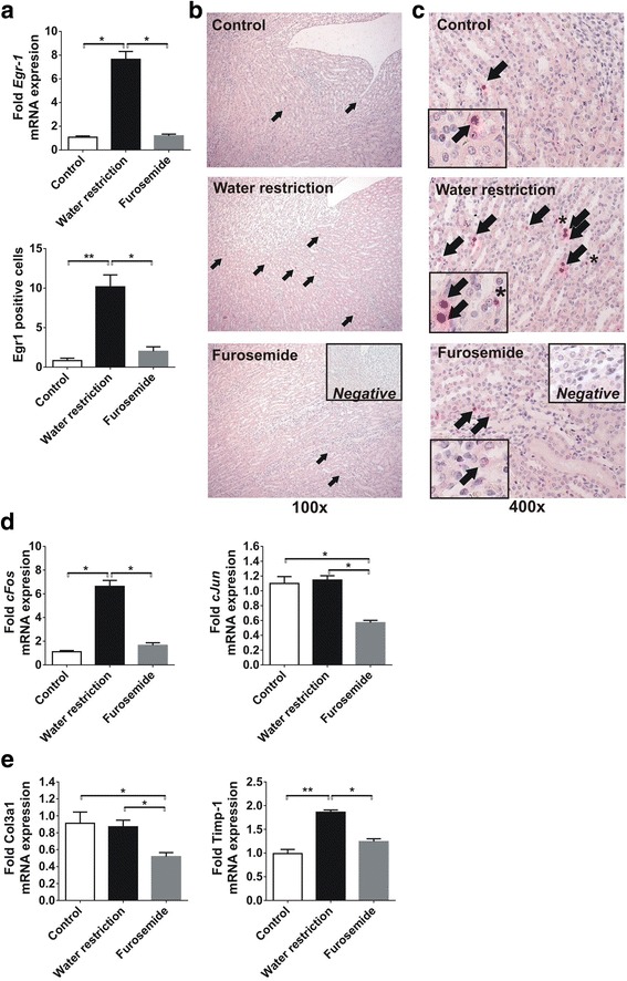
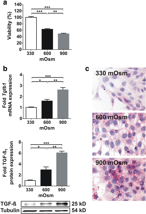
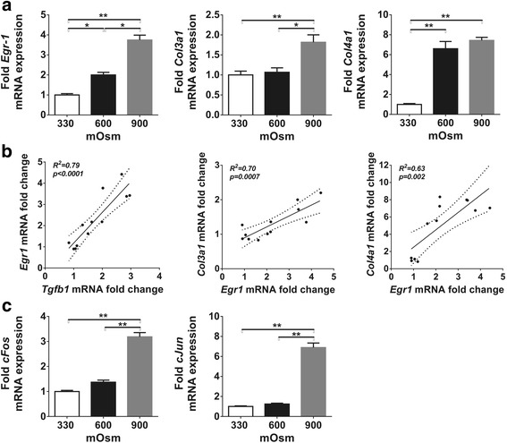
Male adult Sprague Dawley rats (n = 6/group) were treated with 15 mg/day furosemide, or the rats were water restricted to 15 ml/200 g body weight per day. Control rats had free access to water and rodent chow. Kidneys were harvested after 5 days of treament. In cultured inner medullary collecting duct (IMCD) cells, osmolarity was increased from 330 mOsm to 900 mOsm over 6 days. Analyses were performed at 330, 600 and 900 mOsm.
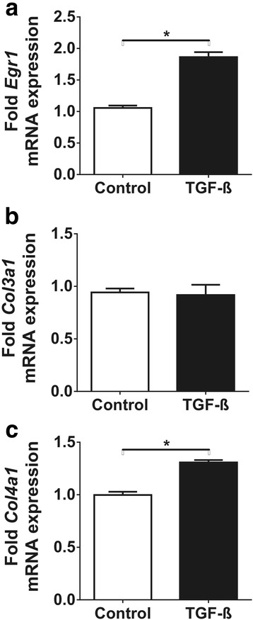
Urine osmolarity has not changed due to furosemide treatment but increased 2-fold after water restriction (p < 0.05). Gene expression of TGF-ß and Egr-1 increased by 1.9-fold and 7-fold in the hypertonic medulla, respectively (p < 0.05), accompanied by 6-fold and 2-fold increased c-Fos and TIMP-1 expression, respectively (p < 0.05) and positive immunostaining for TGF-ß and Egr-1 (p < 0.05). Similarly, hyperosmolarity led to overexpression of TGF-ß and Egr-1 mRNA in IMCD cells (2.5-fold and 3.5-fold increase from 330 to 900 mOsm, respectively (p < 0.05)) accompanied by significant c-Fos and c-Jun overexpressions (p < 0.01), and increased Col3a1 and Col4a1 mRNA expression.
We conclude that both TGF-ß and Egr-1 are upregulated by sustained hyperosmolarity in the rat renal medulla, and it favors the expression of extracellular matrix components.
尽管转化生长因子-β(TGF-β)和转录因子早期生长反应蛋白-1(Egr-1)在肾纤维化及肾髓质渗透压急性变化的反应中均发挥重要作用,但其在持续性低渗或高渗条件下的作用尚未阐明。我们研究了慢性高渗和低渗对肾髓质TGF-β和Egr-1表达的影响。
成年雄性Sprague Dawley大鼠(每组n = 6),每天给予15 mg速尿治疗,或限制大鼠每日饮水量至15 ml/200 g体重。对照大鼠可自由饮水和进食啮齿动物饲料。治疗5天后采集肾脏。在培养的髓质内集合管(IMCD)细胞中,渗透压在6天内从330 mOsm升高至900 mOsm。在330、600和900 mOsm时进行分析。
速尿治疗后尿渗透压未改变,但限水后升高了2倍(p < 0.05)。高渗髓质中TGF-β和Egr-1的基因表达分别增加了1.9倍和7倍(p < 0.05),同时c-Fos和基质金属蛋白酶组织抑制因子-1(TIMP-1)的表达分别增加了6倍和2倍(p < 0.05),且TGF-β和Egr-1免疫染色呈阳性(p < 0.05)。同样,高渗导致IMCD细胞中TGF-β和Egr-1 mRNA过表达(分别从330 mOsm到900 mOsm增加2.5倍和3.5倍,p < 0.05),同时c-Fos和c-Jun显著过表达(p < 0.01),且I型胶原蛋白α1(Col3a1)和IV型胶原蛋白α1(Col4a1)mRNA表达增加。
我们得出结论,大鼠肾髓质中持续性高渗上调了TGF-β和Egr-1的表达,且有利于细胞外基质成分的表达。