• 文献检索
  • 文档翻译
  • 深度研究
  • 学术资讯
  • Suppr Zotero 插件Zotero 插件
  • 邀请有礼
  • 套餐&价格
  • 历史记录
应用&插件
Suppr Zotero 插件Zotero 插件浏览器插件Mac 客户端Windows 客户端微信小程序
定价
高级版会员购买积分包购买API积分包
服务
文献检索文档翻译深度研究API 文档MCP 服务
关于我们
关于 Suppr公司介绍联系我们用户协议隐私条款
关注我们

Suppr 超能文献

核心技术专利:CN118964589B侵权必究
粤ICP备2023148730 号-1Suppr @ 2026

文献检索

告别复杂PubMed语法,用中文像聊天一样搜索,搜遍4000万医学文献。AI智能推荐,让科研检索更轻松。

立即免费搜索

文件翻译

保留排版,准确专业,支持PDF/Word/PPT等文件格式,支持 12+语言互译。

免费翻译文档

深度研究

AI帮你快速写综述,25分钟生成高质量综述,智能提取关键信息,辅助科研写作。

立即免费体验

微孔前处理可改善用于光学和磁共振成像的胰岛标记效果。

Pre-Microporation Improves Outcome of Pancreatic Islet Labelling for Optical and F MR Imaging.

作者信息

Herynek Vít, Gálisová Andrea, Srinivas Mangala, van Dinther Eric A W, Kosinová Lucie, Ruzicka Jiri, Jirátová Markéta, Kriz Jan, Jirák Daniel

机构信息

MR Unit, Radiodiagnostic and Interventional Radiology Department, Institute for Clinical and Experimental Medicine, Vídeňská 1958/9, Prague, Czech Republic.

Department of Tumor Immunology, Radboud University Medical Centre, Route 278, Geert Grooteplein 28, Nijmegen, Netherlands.

出版信息

Biol Proced Online. 2017 Jun 28;19:6. doi: 10.1186/s12575-017-0055-4. eCollection 2017.

DOI:10.1186/s12575-017-0055-4
PMID:28674481
原文链接:https://pmc.ncbi.nlm.nih.gov/articles/PMC5488379/
Abstract

BACKGROUND

In vitro labelling of cells and small cell structures is a necessary step before in vivo monitoring of grafts. We modified and optimised a procedure for pancreatic islet labelling using bimodal positively charged poly(lactic-co-glycolic acid) nanoparticles with encapsulated perfluoro crown ethers and indocyanine green dye via microporation and compared the method with passive endocytosis.

RESULTS

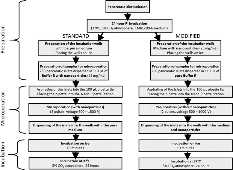
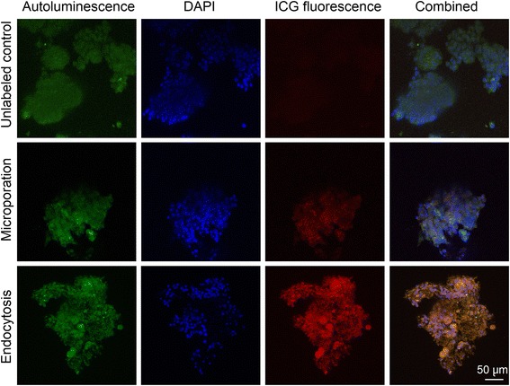
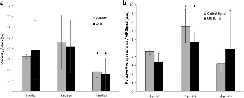
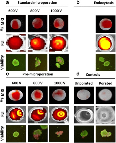
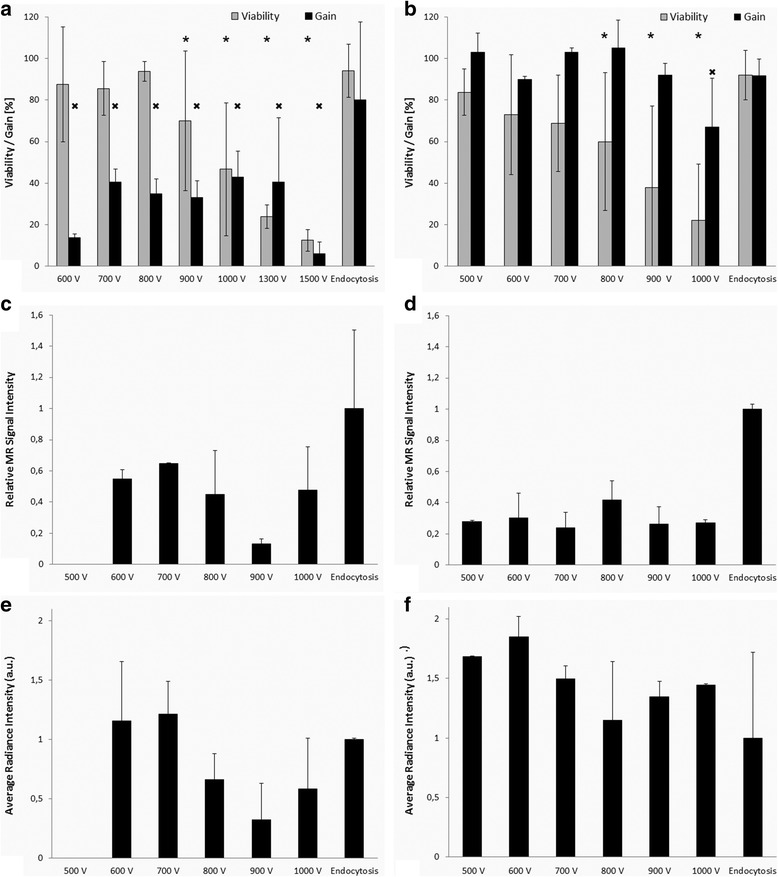
Pancreatic islets were microporated using two pulses at various voltages. We tested a standard procedure (poration in the presence of nanoparticles) and a modified protocol (pre-microporation in a buffer only, and subsequent islet incubation with nanoparticles on ice for 10 min). We compared islet labelling by microporation with labelling by endocytosis, i.e. pancreatic islets were incubated for 24 h in a medium with suspended nanoparticles. In order to verify the efficiency of the labelling procedures, we used F magnetic resonance imaging, optical fluorescence imaging and confocal microscopy. The experiment confirmed that microporation, albeit fast and effective, is invasive and may cause substantial harm to islets. To achieve sufficient poration and to minimise the reduction of viability, the electric field should be set at 20 kV/m (two pulses, 20 ms each). Poration in the presence of nanoparticles was found to be unsuitable for the nanoparticles used. The water suspension of nanoparticles (which served as a surfactant) was slightly foamy and microbubbles in the suspension were responsible for sparks causing the destruction of islets during poration. However, pre-microporation (poration of islets in a buffer only) followed by 10-min incubation with nanoparticles was safer.

CONCLUSIONS

For labelling of pancreatic islets using poly(lactic-co-glycolic acid) nanoparticles, the modified microporation procedure with low voltage was found to be safer than the standard microporation procedure. The modified procedure was fast, however, efficiency was lower compared to endocytosis.

摘要

背景

在对移植体进行体内监测之前,对细胞和小细胞结构进行体外标记是必要步骤。我们改进并优化了一种使用双模态带正电荷的聚乳酸-乙醇酸共聚物纳米颗粒(其包裹有全氟冠醚和吲哚菁绿染料)通过微孔法对胰岛进行标记的程序,并将该方法与被动内吞作用进行了比较。

结果

使用不同电压的两个脉冲对胰岛进行微孔处理。我们测试了一种标准程序(在纳米颗粒存在下进行微孔处理)和一种改进方案(仅在缓冲液中进行预微孔处理,随后将胰岛与纳米颗粒在冰上孵育10分钟)。我们将通过微孔法进行的胰岛标记与通过内吞作用进行的标记进行了比较,即把胰岛在含有悬浮纳米颗粒的培养基中孵育24小时。为了验证标记程序的效率,我们使用了F磁共振成像、光学荧光成像和共聚焦显微镜。实验证实,尽管微孔法快速有效,但具有侵入性,可能对胰岛造成实质性损害。为了实现足够的微孔处理并将活力降低最小化,电场应设置为20 kV/m(两个脉冲,每个脉冲20毫秒)。发现在纳米颗粒存在下进行微孔处理不适用于所使用的纳米颗粒。纳米颗粒的水悬浮液(用作表面活性剂)略有泡沫,悬浮液中的微气泡导致在微孔处理期间产生火花,从而破坏胰岛。然而,先进行预微孔处理(仅在缓冲液中对胰岛进行微孔处理),然后与纳米颗粒孵育10分钟则更安全。

结论

对于使用聚乳酸-乙醇酸共聚物纳米颗粒对胰岛进行标记,发现低电压的改进微孔处理程序比标准微孔处理程序更安全。改进后的程序速度快,然而,与内吞作用相比效率较低。

https://cdn.ncbi.nlm.nih.gov/pmc/blobs/59e3/5488379/2ae2752c7110/12575_2017_55_Fig6_HTML.jpg
https://cdn.ncbi.nlm.nih.gov/pmc/blobs/59e3/5488379/e27fb243f7c8/12575_2017_55_Fig1_HTML.jpg
https://cdn.ncbi.nlm.nih.gov/pmc/blobs/59e3/5488379/47248a19f052/12575_2017_55_Fig2_HTML.jpg
https://cdn.ncbi.nlm.nih.gov/pmc/blobs/59e3/5488379/be8f3d720027/12575_2017_55_Fig3_HTML.jpg
https://cdn.ncbi.nlm.nih.gov/pmc/blobs/59e3/5488379/e8935b667e3a/12575_2017_55_Fig4_HTML.jpg
https://cdn.ncbi.nlm.nih.gov/pmc/blobs/59e3/5488379/4c3382ed4a7b/12575_2017_55_Fig5_HTML.jpg
https://cdn.ncbi.nlm.nih.gov/pmc/blobs/59e3/5488379/2ae2752c7110/12575_2017_55_Fig6_HTML.jpg

相似文献

1
Pre-Microporation Improves Outcome of Pancreatic Islet Labelling for Optical and F MR Imaging.微孔前处理可改善用于光学和磁共振成像的胰岛标记效果。
Biol Proced Online. 2017 Jun 28;19:6. doi: 10.1186/s12575-017-0055-4. eCollection 2017.
2
A Trimodal Imaging Platform for Tracking Viable Transplanted Pancreatic Islets In Vivo: F-19 MR, Fluorescence, and Bioluminescence Imaging.一种用于在体追踪可存活移植胰岛的三模态成像平台:F-19MR、荧光和生物发光成像。
Mol Imaging Biol. 2019 Jun;21(3):454-464. doi: 10.1007/s11307-018-1270-3.
3
In vivo imaging of islet transplantation using PLGA nanoparticles containing iron oxide and indocyanine green.使用含有氧化铁和吲哚菁绿的聚乳酸-羟基乙酸共聚物纳米颗粒对胰岛移植进行体内成像。
Magn Reson Med. 2014 Mar;71(3):1054-63. doi: 10.1002/mrm.24768.
4
In Vivo Magnetic Resonance Imaging of Small Interfering RNA Nanodelivery to Pancreatic Islets.小干扰RNA纳米递送至胰岛的体内磁共振成像
Methods Mol Biol. 2016;1372:25-36. doi: 10.1007/978-1-4939-3148-4_2.
5
Construction of functional pancreatic artificial islet tissue composed of fibroblast-modified polylactic- co-glycolic acid membrane and pancreatic stem cells.由成纤维细胞修饰的聚乳酸-乙醇酸共聚物膜和胰腺干细胞构建功能性胰腺人工胰岛组织
J Biomater Appl. 2017 Sep;32(3):362-372. doi: 10.1177/0885328217722041. Epub 2017 Jul 26.
6
MRI of transplanted surface-labeled pancreatic islets with heparinized superparamagnetic iron oxide nanoparticles.用肝素化超顺磁氧化铁纳米颗粒对移植的表面标记胰岛进行 MRI。
Biomaterials. 2011 Dec;32(35):9391-400. doi: 10.1016/j.biomaterials.2011.08.070. Epub 2011 Sep 8.
7
Magnetic resonance imaging and biological properties of pancreatic islets labeled with iron oxide nanoparticles.氧化铁纳米颗粒标记的胰岛的磁共振成像及生物学特性
NMR Biomed. 2009 Oct;22(8):852-6. doi: 10.1002/nbm.1398.
8
Effects of glucose toxicity and islet purity on in vivo magnetic resonance imaging of transplanted pancreatic islets.葡萄糖毒性和胰岛纯度对移植胰岛体内磁共振成像的影响。
Transplantation. 2008 Apr 27;85(8):1091-8. doi: 10.1097/TP.0b013e31816b183e.
9
Tri-modal In vivo Imaging of Pancreatic Islets Transplanted Subcutaneously in Mice.三模态活体成像监测小鼠皮下移植胰岛。
Mol Imaging Biol. 2018 Dec;20(6):940-951. doi: 10.1007/s11307-018-1192-0.
10
Evidence of pituitary adenylate cyclase activating polypeptide (PACAP) in pancreatic islet cells by confocal microscopy.通过共聚焦显微镜观察胰腺胰岛细胞中垂体腺苷酸环化酶激活多肽(PACAP)的证据。
Pancreas. 2001 Jul;23(1):68-71. doi: 10.1097/00006676-200107000-00010.

引用本文的文献

1
Cationic fluorinated micelles for cell labeling and F-MR imaging.阳离子氟化物胶束用于细胞标记和 F-MR 成像。
Sci Rep. 2024 Sep 30;14(1):22613. doi: 10.1038/s41598-024-73511-8.
2
Imaging of Pancreatic Islet Grafts in Diabetes Treatment.胰岛移植治疗糖尿病的影像学评估
Front Endocrinol (Lausanne). 2021 Mar 2;12:640117. doi: 10.3389/fendo.2021.640117. eCollection 2021.
3
Islet Transplantation Imaging in vivo.体内胰岛移植成像

本文引用的文献

1
In vivo and ex vivo 19-fluorine magnetic resonance imaging and spectroscopy of beta-cells and pancreatic islets using GLUT-2 specific contrast agents.使用GLUT-2特异性造影剂对β细胞和胰岛进行体内和体外19氟磁共振成像及波谱分析。
Contrast Media Mol Imaging. 2016 Nov;11(6):506-513. doi: 10.1002/cmmi.1712. Epub 2016 Sep 14.
2
Tracking the fate of stem cell implants with fluorine-19 MRI.利用氟-19磁共振成像追踪干细胞植入物的命运。
PLoS One. 2015 Mar 13;10(3):e0118544. doi: 10.1371/journal.pone.0118544. eCollection 2015.
3
Cell tracking using (19)F magnetic resonance imaging: technical aspects and challenges towards clinical applications.
Diabetes Metab Syndr Obes. 2020 Sep 23;13:3301-3311. doi: 10.2147/DMSO.S263253. eCollection 2020.
4
A Trimodal Imaging Platform for Tracking Viable Transplanted Pancreatic Islets In Vivo: F-19 MR, Fluorescence, and Bioluminescence Imaging.一种用于在体追踪可存活移植胰岛的三模态成像平台:F-19MR、荧光和生物发光成像。
Mol Imaging Biol. 2019 Jun;21(3):454-464. doi: 10.1007/s11307-018-1270-3.
使用 (19)F 磁共振成像进行细胞示踪:技术方面及向临床应用的挑战。
Eur Radiol. 2015 Mar;25(3):726-35. doi: 10.1007/s00330-014-3474-5. Epub 2014 Nov 6.
4
In vivo MR detection of fluorine-labeled human MSC using the bSSFP sequence.利用 bSSFP 序列在体检测氟标记的人 MSC。
Int J Nanomedicine. 2014 Apr 8;9:1731-9. doi: 10.2147/IJN.S59127. eCollection 2014.
5
Combining perfluorocarbon and superparamagnetic iron-oxide cell labeling for improved and expanded applications of cellular MRI.结合全氟化碳和超顺磁性氧化铁细胞标记以改进和拓展细胞磁共振成像的应用
Magn Reson Med. 2015 Jan;73(1):367-75. doi: 10.1002/mrm.25120. Epub 2014 Jan 29.
6
Tracking immune cells in vivo using magnetic resonance imaging.利用磁共振成像技术在体内追踪免疫细胞。
Nat Rev Immunol. 2013 Oct;13(10):755-63. doi: 10.1038/nri3531. Epub 2013 Sep 10.
7
Seeing stem cells at work in vivo.在体内观察干细胞的工作情况。
Stem Cell Rev Rep. 2014 Feb;10(1):127-44. doi: 10.1007/s12015-013-9468-x.
8
Microfluidic electroporation for cellular analysis and delivery.微流控电穿孔用于细胞分析和药物递送。
Lab Chip. 2013 Oct 7;13(19):3803-21. doi: 10.1039/c3lc50566a.
9
In vivo imaging of transplanted islets labeled with a novel cationic nanoparticle.新型阳离子纳米颗粒标记的移植胰岛的体内成像。
PLoS One. 2013;8(2):e57046. doi: 10.1371/journal.pone.0057046. Epub 2013 Feb 22.
10
The role of surface charge in cellular uptake and cytotoxicity of medical nanoparticles.医学纳米粒子表面电荷在细胞摄取和细胞毒性中的作用。
Int J Nanomedicine. 2012;7:5577-91. doi: 10.2147/IJN.S36111. Epub 2012 Nov 2.