Erskine Lynda, François Urielle, Denti Laura, Joyce Andy, Tillo Miguel, Bruce Freyja, Vargesson Neil, Ruhrberg Christiana
School of Medicine, Medical Sciences and Nutrition, Institute of Medical Sciences, University of Aberdeen, Aberdeen AB25 2ZD, UK
School of Medicine, Medical Sciences and Nutrition, Institute of Medical Sciences, University of Aberdeen, Aberdeen AB25 2ZD, UK.
Development. 2017 Jul 1;144(13):2504-2516. doi: 10.1242/dev.151621.
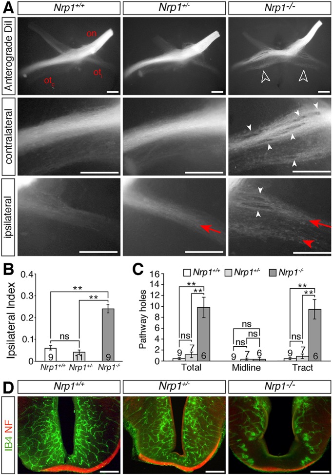
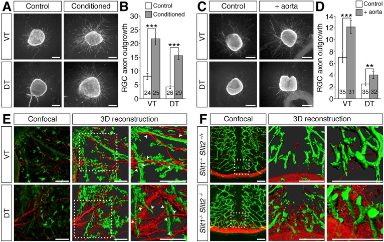
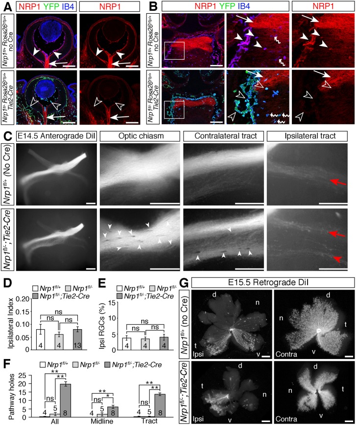
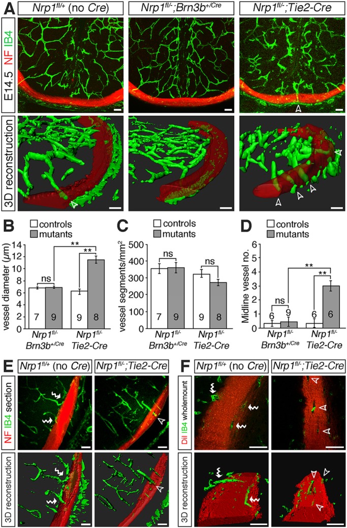
Visual information is relayed from the eye to the brain via retinal ganglion cell (RGC) axons. Mice lacking NRP1 or NRP1-binding VEGF-A isoforms have defective RGC axon organisation alongside brain vascular defects. It is not known whether axonal defects are caused exclusively by defective VEGF-A signalling in RGCs or are exacerbated by abnormal vascular morphology. Targeted NRP1 ablation in RGCs with a knock-in allele reduced axonal midline crossing at the optic chiasm and optic tract fasciculation. In contrast, -mediated endothelial NRP1 ablation induced axon exclusion zones in the optic tracts without impairing axon crossing. Similar defects were observed in and mice, which have vascular defects as a result of their expression of single VEGF-A isoforms. Ectopic midline vascularisation in endothelial and mutants caused additional axonal exclusion zones within the chiasm. As and assays demonstrated that vessels do not repel axons, abnormally large or ectopically positioned vessels are likely to present physical obstacles to axon growth. We conclude that proper axonal wiring during brain development depends on the precise molecular control of neurovascular co-patterning.
视觉信息通过视网膜神经节细胞(RGC)轴突从眼睛传递至大脑。缺乏NRP1或与NRP1结合的VEGF - A亚型的小鼠,其RGC轴突组织存在缺陷,同时伴有脑血管缺陷。目前尚不清楚轴突缺陷是否仅由RGC中VEGF - A信号传导缺陷引起,还是由异常的血管形态加剧。利用敲入等位基因在RGC中靶向消融NRP1,减少了视交叉处轴突的中线交叉和视束束化。相比之下,介导的内皮细胞NRP1消融在视束中诱导了轴突排斥区,但不影响轴突交叉。在 和 小鼠中也观察到类似的缺陷,它们由于表达单一VEGF - A亚型而存在血管缺陷。内皮细胞 和 突变体中的异位中线血管生成在视交叉内导致了额外的轴突排斥区。由于 和 实验表明血管不会排斥轴突,异常大或异位定位的血管可能会对轴突生长造成物理障碍。我们得出结论,大脑发育过程中正确的轴突布线取决于神经血管共同模式形成的精确分子控制。