van Gijtenbeek Lieke A, Kok Jan
Department of Molecular Genetics, Faculty of Science and Engineering, University of GroningenGroningen, Netherlands.
Front Microbiol. 2017 Jun 22;8:1161. doi: 10.3389/fmicb.2017.01161. eCollection 2017.
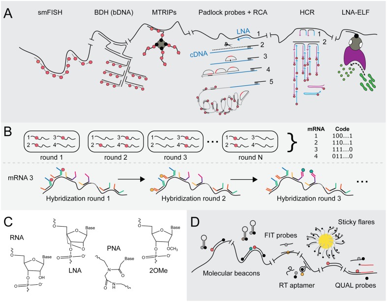
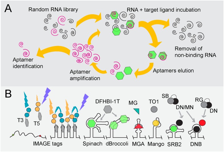
To be able to visualize the abundance and spatiotemporal features of RNAs in bacterial cells would permit obtaining a pivotal understanding of many mechanisms underlying bacterial cell biology. The first methods that allowed observing single mRNA molecules in individual cells were introduced by Bertrand et al. (1998) and Femino et al. (1998). Since then, a plethora of techniques to image RNA molecules with the aid of fluorescence microscopy has emerged. Many of these approaches are useful for the large eukaryotic cells but their adaptation to study RNA, specifically mRNA molecules, in bacterial cells progressed relatively slow. Here, an overview will be given of fluorescent techniques that can be used to reveal specific RNA molecules inside fixed and living single bacterial cells. It includes a critical evaluation of their caveats as well as potential solutions.
能够可视化细菌细胞中RNA的丰度和时空特征,将有助于对细菌细胞生物学的许多潜在机制获得关键的理解。Bertrand等人(1998年)和Femino等人(1998年)首次引入了能够在单个细胞中观察单个mRNA分子的方法。从那时起,借助荧光显微镜对RNA分子进行成像的大量技术应运而生。其中许多方法对大型真核细胞很有用,但它们在细菌细胞中用于研究RNA,特别是mRNA分子的适应性进展相对缓慢。本文将概述可用于揭示固定和活的单个细菌细胞内特定RNA分子的荧光技术。它包括对其缺点以及潜在解决方案的批判性评估。