Sadegh Sanaz, Higgins Jenny L, Mannion Patrick C, Tamkun Michael M, Krapf Diego
Department of Electrical and Computer Engineering, Colorado State University, Fort Collins, Colorado 80523, USA.
School of Biomedical Engineering, Colorado State University, Fort Collins, Colorado 80523, USA.
Phys Rev X. 2017 Jan-Mar;7(1). doi: 10.1103/PhysRevX.7.011031. Epub 2017 Mar 9.
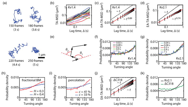
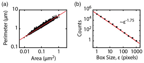
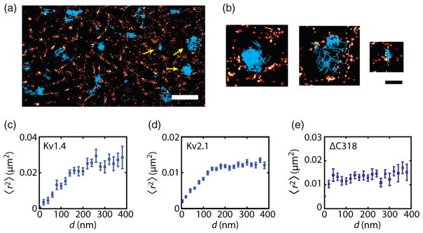
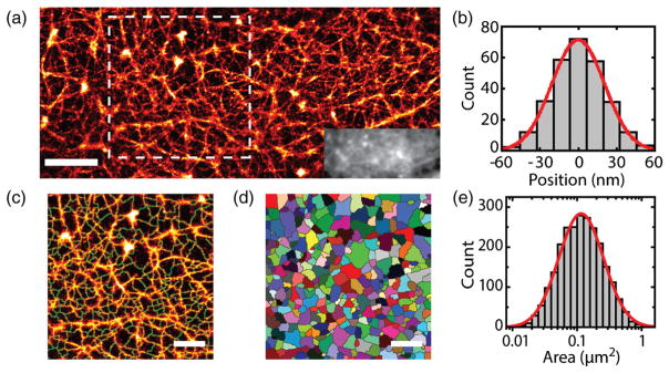
A broad range of membrane proteins display anomalous diffusion on the cell surface. Different methods provide evidence for obstructed subdiffusion and diffusion on a fractal space, but the underlying structure inducing anomalous diffusion has never been visualized because of experimental challenges. We addressed this problem by imaging the cortical actin at high resolution while simultaneously tracking individual membrane proteins in live mammalian cells. Our data confirm that actin introduces barriers leading to compartmentalization of the plasma membrane and that membrane proteins are transiently confined within actin fences. Furthermore, superresolution imaging shows that the cortical actin is organized into a self-similar meshwork. These results present a hierarchical nanoscale picture of the plasma membrane.
多种膜蛋白在细胞表面呈现异常扩散。不同方法为受阻的亚扩散和分形空间上的扩散提供了证据,但由于实验挑战,导致异常扩散的潜在结构从未被可视化。我们通过在高分辨率下对皮层肌动蛋白成像,同时在活的哺乳动物细胞中追踪单个膜蛋白来解决这个问题。我们的数据证实,肌动蛋白引入了屏障,导致质膜分隔,并且膜蛋白被短暂限制在肌动蛋白围栏内。此外,超分辨率成像显示皮层肌动蛋白组织成自相似网络。这些结果呈现了质膜的分级纳米尺度图景。