Bolognesi Maddalena Maria, Manzoni Marco, Scalia Carla Rossana, Zannella Stefano, Bosisio Francesca Maria, Faretta Mario, Cattoretti Giorgio
Dipartimento di Medicina e Chirurgia, Universitá degli Studi di Milano-Bicocca, Monza, Italy (MMB, MM, CRS, SZ, FMB, GC).
Laboratory of Translational Cell and Tissue Research, KU Leuven, Leuven, Belgium (FMB).
J Histochem Cytochem. 2017 Aug;65(8):431-444. doi: 10.1369/0022155417719419. Epub 2017 Jul 10.
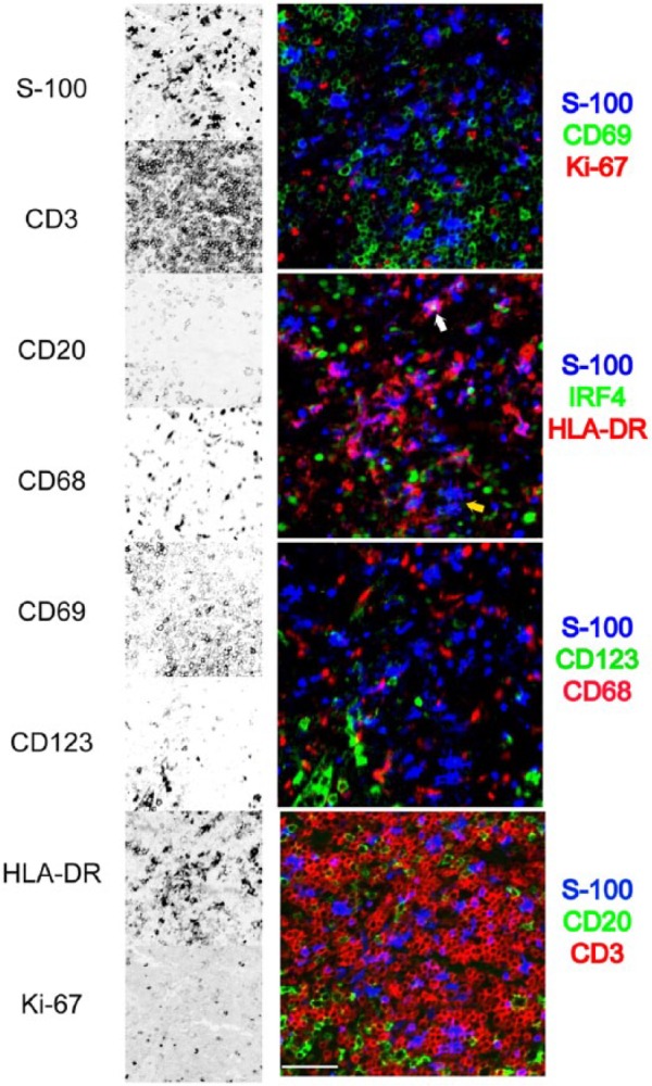
Multiplexing, labeling for multiple immunostains in the very same cell or tissue section in situ, has raised considerable interest. The methods proposed include the use of labeled primary antibodies, spectral separation of fluorochromes, bleaching of the fluorophores or chromogens, blocking of previous antibody layers, all in various combinations. The major obstacles to the diffusion of this technique are high costs in custom antibodies and instruments, low throughput, and scarcity of specialized skills or facilities. We have validated a method based on common primary and secondary antibodies and diffusely available fluorescent image scanners. It entails rounds of four-color indirect immunofluorescence, image acquisition, and removal (stripping) of the antibodies, before another stain is applied. The images are digitally registered and the autofluorescence is subtracted. Removal of antibodies is accomplished by disulfide cleavage and a detergent or by a chaotropic salt treatment, this latter followed by antigen refolding. More than 30 different antibody stains can be applied to one single section from routinely fixed and embedded tissue. This method requires a modest investment in hardware and materials and uses freeware image analysis software. Multiplexing on routine tissue sections is a high throughput tool for in situ characterization of neoplastic, reactive, inflammatory, and normal cells.
多重标记,即在同一细胞或组织切片原位进行多种免疫染色标记,已经引起了广泛关注。提出的方法包括使用标记的一抗、荧光染料的光谱分离、荧光团或色原的漂白、阻断先前的抗体层,所有这些方法都有不同的组合。这项技术推广的主要障碍是定制抗体和仪器成本高、通量低以及缺乏专业技能或设施。我们验证了一种基于常见一抗和二抗以及普遍可用的荧光图像扫描仪的方法。该方法包括进行多轮四色间接免疫荧光、图像采集以及在施加另一种染色之前去除(剥离)抗体。图像进行数字配准并减去自发荧光。通过二硫键裂解和去污剂或通过离液盐处理来去除抗体,后者之后进行抗原重折叠。可以将30多种不同的抗体染色应用于常规固定和包埋组织的单个切片。该方法在硬件和材料方面需要适度投资,并使用免费的图像分析软件。对常规组织切片进行多重标记是一种用于原位表征肿瘤细胞、反应性细胞、炎症细胞和正常细胞的高通量工具。