• 文献检索
  • 文档翻译
  • 深度研究
  • 学术资讯
  • Suppr Zotero 插件Zotero 插件
  • 邀请有礼
  • 套餐&价格
  • 历史记录
应用&插件
Suppr Zotero 插件Zotero 插件浏览器插件Mac 客户端Windows 客户端微信小程序
定价
高级版会员购买积分包购买API积分包
服务
文献检索文档翻译深度研究API 文档MCP 服务
关于我们
关于 Suppr公司介绍联系我们用户协议隐私条款
关注我们

Suppr 超能文献

核心技术专利:CN118964589B侵权必究
粤ICP备2023148730 号-1Suppr @ 2026

文献检索

告别复杂PubMed语法,用中文像聊天一样搜索,搜遍4000万医学文献。AI智能推荐,让科研检索更轻松。

立即免费搜索

文件翻译

保留排版,准确专业,支持PDF/Word/PPT等文件格式,支持 12+语言互译。

免费翻译文档

深度研究

AI帮你快速写综述,25分钟生成高质量综述,智能提取关键信息,辅助科研写作。

立即免费体验

生化和电子显微镜证据表明,在海胆早期发育过程中,细胞核对线粒体基因组活性起负向控制作用。

Biochemical and electron microscopic evidence that cell nucleus negatively controls mitochondrial genomic activity in early sea urchin development.

作者信息

Rinaldi A M, De Leo G, Arzone A, Salcher I, Storace A, Mutolo V

出版信息

Proc Natl Acad Sci U S A. 1979 Apr;76(4):1916-20. doi: 10.1073/pnas.76.4.1916.

DOI:10.1073/pnas.76.4.1916
PMID:287031
原文链接:https://pmc.ncbi.nlm.nih.gov/articles/PMC383503/
Abstract

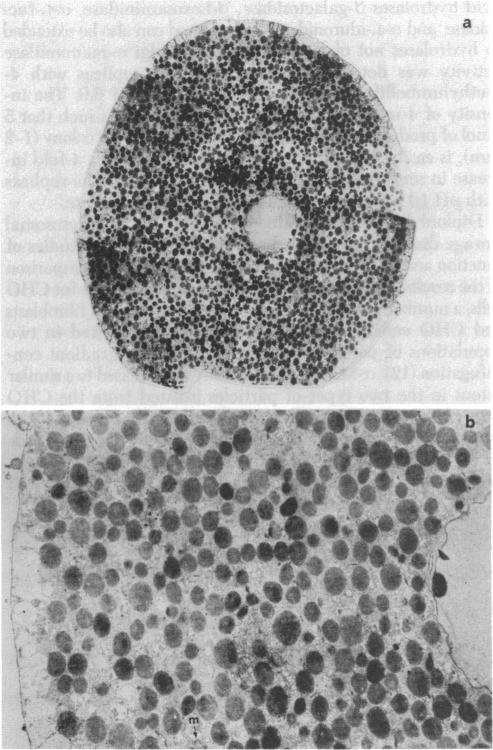
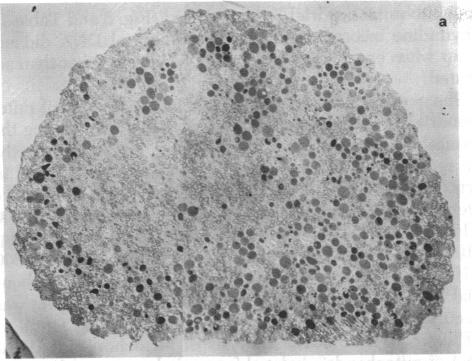
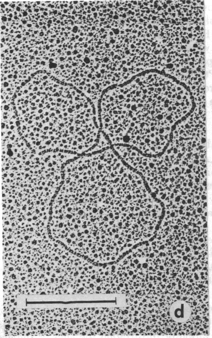
Enucleated halves of sea urchin eggs obtained by centrifugation contain almost all the mitochondrial population of the egg. Removal of the nucleus followed by parthenogenetic activation stimulates the incorporation of [3H]thymidine into the mitochondrial DNA, whereas no such incorportion is observed in activated whole eggs. The block is not the result of a modification in the permeability of the mitochondrial membrane. Electron microscopic observations demonstrated duplication of mitochondrial DNA molecules in activated enucleated halves. No duplication was found in the mitochondrial DNA from activated whole eggs or from nonactivated enucleated halves. We conclude that the cell nucleus exerts a negative control on the activity of the mitochondrial genome through some short-lived nuclear substance(s).

摘要

通过离心获得的海胆卵去核半卵包含了卵中几乎所有的线粒体群体。去除细胞核后进行孤雌激活会刺激[3H]胸腺嘧啶核苷掺入线粒体DNA,而在激活的完整卵中未观察到这种掺入。这种阻断不是线粒体膜通透性改变的结果。电子显微镜观察表明,激活的去核半卵中线粒体DNA分子发生了复制。在激活的完整卵或未激活的去核半卵的线粒体DNA中未发现复制现象。我们得出结论,细胞核通过某种短命的核物质对线粒体基因组的活性施加负调控。

https://cdn.ncbi.nlm.nih.gov/pmc/blobs/ab01/383503/4b9d9404ca23/pnas00004-0401-c.jpg
https://cdn.ncbi.nlm.nih.gov/pmc/blobs/ab01/383503/0674b44e5b3e/pnas00004-0398-a.jpg
https://cdn.ncbi.nlm.nih.gov/pmc/blobs/ab01/383503/5f2a52fecb83/pnas00004-0399-a.jpg
https://cdn.ncbi.nlm.nih.gov/pmc/blobs/ab01/383503/f211c11c5505/pnas00004-0399-b.jpg
https://cdn.ncbi.nlm.nih.gov/pmc/blobs/ab01/383503/00f47f3a2666/pnas00004-0400-a.jpg
https://cdn.ncbi.nlm.nih.gov/pmc/blobs/ab01/383503/fded3ba0c244/pnas00004-0400-b.jpg
https://cdn.ncbi.nlm.nih.gov/pmc/blobs/ab01/383503/444707a9c3d6/pnas00004-0400-c.jpg
https://cdn.ncbi.nlm.nih.gov/pmc/blobs/ab01/383503/b68526165f49/pnas00004-0400-d.jpg
https://cdn.ncbi.nlm.nih.gov/pmc/blobs/ab01/383503/8afc2d1cd7e8/pnas00004-0400-e.jpg
https://cdn.ncbi.nlm.nih.gov/pmc/blobs/ab01/383503/c3cd33f4135d/pnas00004-0400-f.jpg
https://cdn.ncbi.nlm.nih.gov/pmc/blobs/ab01/383503/565854e847b0/pnas00004-0400-g.jpg
https://cdn.ncbi.nlm.nih.gov/pmc/blobs/ab01/383503/3ab8ffa6e544/pnas00004-0401-a.jpg
https://cdn.ncbi.nlm.nih.gov/pmc/blobs/ab01/383503/c2d2e43915ff/pnas00004-0401-b.jpg
https://cdn.ncbi.nlm.nih.gov/pmc/blobs/ab01/383503/4b9d9404ca23/pnas00004-0401-c.jpg

相似文献

1
Biochemical and electron microscopic evidence that cell nucleus negatively controls mitochondrial genomic activity in early sea urchin development.生化和电子显微镜证据表明,在海胆早期发育过程中,细胞核对线粒体基因组活性起负向控制作用。
Proc Natl Acad Sci U S A. 1979 Apr;76(4):1916-20. doi: 10.1073/pnas.76.4.1916.
2
Studies of the mechanisms of nuclear control over the synthesis of mitochondrial DNA in sea urchin eggs.海胆卵细胞核对线粒体DNA合成的控制机制研究。
Cell Biol Int Rep. 1989 Feb;13(2):181-7. doi: 10.1016/0309-1651(89)90064-7.
3
Mitochondrial division in non nucleated sea urchin eggs.无核海胆卵中的线粒体分裂
Cell Biol Int Rep. 1979 Mar;3(2):179-82. doi: 10.1016/0309-1651(79)90124-3.
4
[In vivo synthesis of mitochondrial DNA during embryonal development of Paracentrotus lividus].[紫海胆胚胎发育过程中线粒体DNA的体内合成]
Boll Soc Ital Biol Sper. 1980 May 30;56(10):1024-30.
5
The nucleus negatively controls the synthesis of mitochondrial proteins in the sea urchin egg.
Cell Biol Int Rep. 1983 Mar;7(3):211-8. doi: 10.1016/0309-1651(83)90228-x.
6
Mitochondrial DNA replication in sea urchin oocytes.海胆卵母细胞中的线粒体DNA复制
J Cell Biol. 1974 Oct;63(1):146-59. doi: 10.1083/jcb.63.1.146.
7
Heterogeneous distribution and replication activity of mitochondria in Xenopus laevis oocytes.
Eur J Cell Biol. 1984 May;34(1):171-8.
8
Relationship between nuclear DNA synthesis and centrosome reproduction in sea urchin eggs.海胆卵中核DNA合成与中心体复制之间的关系。
J Exp Zool. 1987 Oct;244(1):89-100. doi: 10.1002/jez.1402440111.
9
Localization of 3H-thymidine in oocyte Mitochondria from Schistosoma mansoni.3H-胸腺嘧啶在曼氏血吸虫卵母细胞线粒体中的定位
J Parasitol. 1981 Jun;67(3):355-61.
10
Postfertilization growth of microvilli in the sea urchin egg: new views from eggs that have been quick-frozen, freeze-fractured, and deeply etched.海胆卵受精后微绒毛的生长:来自快速冷冻、冷冻断裂和深度蚀刻卵的新观点。
Dev Biol. 1981 Mar;82(2):393-400. doi: 10.1016/0012-1606(81)90463-2.

引用本文的文献

1
Mitochondrial growth during the cell cycle of Trypanosoma brucei bloodstream forms.布氏锥虫血流形式细胞周期中的线粒体生长
Sci Rep. 2016 Nov 22;6:36565. doi: 10.1038/srep36565.
2
A method for measuring mitochondrial mass and activity.一种测量线粒体质量和活性的方法。
Cytotechnology. 2008 Mar;56(3):145-9. doi: 10.1007/s10616-008-9143-2. Epub 2008 Jun 24.
3
The posterior determinant gene nanos is required for the maintenance of the adult germline stem cells during Drosophila oogenesis.在后生动物果蝇卵子发生过程中,后决定基因nanos对于成体生殖系干细胞的维持是必需的。

本文引用的文献

1
The use of lead citrate at high pH as an electron-opaque stain in electron microscopy.在电子显微镜检查中,将高pH值的柠檬酸铅用作电子不透明染色剂。
J Cell Biol. 1963 Apr;17(1):208-12. doi: 10.1083/jcb.17.1.208.
2
Synthesis of ribosomal RNA during sea-urchin development.海胆发育过程中核糖体RNA的合成
Biochim Biophys Acta. 1967 Apr 18;138(2):276-85. doi: 10.1016/0005-2787(67)90488-1.
3
Transcription of nuclear and cytoplasmic genes during early development of sea urchin embryos.
Genetics. 1999 Apr;151(4):1479-92. doi: 10.1093/genetics/151.4.1479.
4
Sea urchin egg mitochondrial DNA contains a short displacement loop (D-loop) in the replication origin region.海胆卵线粒体DNA在复制起始区域包含一个短的置换环(D环)。
Nucleic Acids Res. 1989 Nov 25;17(22):8949-65. doi: 10.1093/nar/17.22.8949.
J Mol Biol. 1969 Apr 14;41(1):155-8. doi: 10.1016/0022-2836(69)90133-8.
4
Sea urchin embryogenesis. I. RNA synthesis by cytoplasmic and nuclear genes during development.
Dev Biol. 1971 Jun;25(2):209-31. doi: 10.1016/0012-1606(71)90028-5.
5
RNA synthesis in anucleate egg fragments and normal embryos of the sea urchin, Arbacia punctulata.
Biochim Biophys Acta. 1970 Jul 16;213(1):183-93. doi: 10.1016/0005-2787(70)90019-5.
6
Synthesis of RNA in non-nucleate fragments of sea urchin eggs.
J Mol Biol. 1970 Feb 14;47(3):615-8. doi: 10.1016/0022-2836(70)90331-1.
7
Protein synthesis and development in the absence of cytoplasmic RNA synthesis in nonnucleate egg fragments and embryos of sea urchins: effect of ethidium bromide.
Dev Biol. 1971 Feb;24(2):214-32. doi: 10.1016/0012-1606(71)90096-0.
8
Mitochondrial RNA synthesis in sea urchin embryos.海胆胚胎中的线粒体RNA合成。
J Mol Biol. 1972 Mar 14;64(3):593-607. doi: 10.1016/0022-2836(72)90085-x.
9
Cytoplasmic synthesis of RNA in the sea urchin embryo. II. Mitochondrial transcription.
Cell Differ. 1972 Apr;1(1):5-14. doi: 10.1016/0045-6039(72)90107-8.
10
An analysis of the partial metabolic derepression of sea urchin eggs by ammonia: the existence of independent pathways.氨对海胆卵部分代谢去抑制作用的分析:独立途径的存在
Dev Biol. 1974 Oct;40(2):245-55. doi: 10.1016/0012-1606(74)90127-4.