OMFS-IMPATH Research Group, Department of Imaging and Pathology, Faculty of Medicine, Catholic University Leuven, Leuven, Belgium.
Oral and Maxillofacial Surgery Department, Faculty of Dentistry, Mansoura University, Mansoura, Egypt.
Sci Rep. 2017 Jul 13;7(1):5356. doi: 10.1038/s41598-017-05788-x.
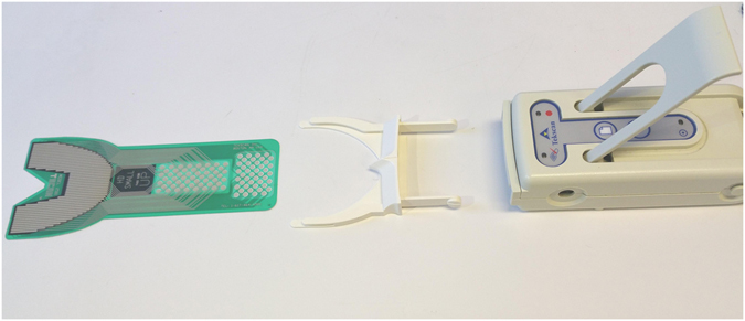
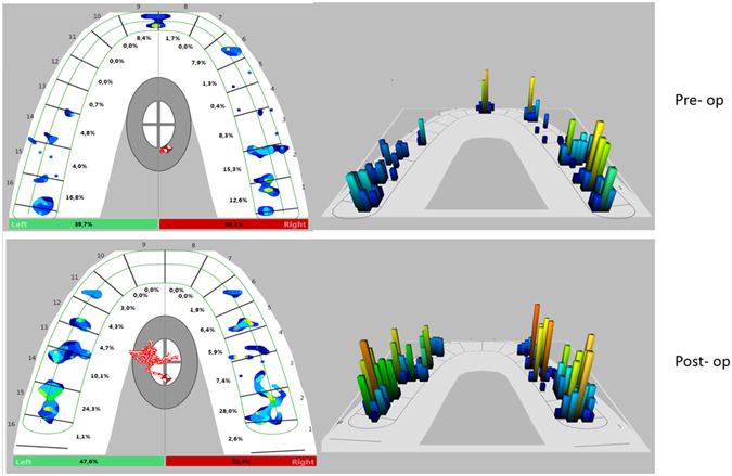
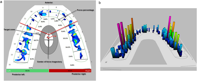
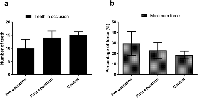
Dental occlusion varies among individuals, and achieving correct physiological occlusion after osteotomy is essential for the complex functioning of the stomatognathic system. The T-Scan system records the centre of force, first contact, maximum bite force, and maximum intercuspation. The aim of the present study was to investigate the usefulness and consistency of T-Scan in assessing occlusion before and after orthognathic surgery. Occlusal information was evaluated for 30 healthy adults with normal occlusion and 40 patients undergoing orthognathic surgery. T-Scan had a high degree of reliability for consecutive measurements (Pearson correlation, r = 0.98). For most parameters, occlusal distribution was better after surgery than before surgery. More teeth contributed to occlusion at maximum intercuspation after surgery than before surgery (14 vs. 10). In addition, the difference in the posterior force distribution was reduced after surgery (17.6 ± 13.8 vs. 22.7 ± 21.4 before surgery), indicating better occlusal force distribution after surgery. The maximum percentage force on teeth (p = 0.004) and the number of teeth contributing to occlusion (p < 0.001) also differed significantly. Thus, T-Scan is good for assessing occlusal discrepancies and can be used to portray the pre- and post-operative occlusal contact distribution during treatment planning and follow-up.
个体间的咬合存在差异,而在截骨术后实现正确的生理咬合对于咀嚼系统的复杂功能至关重要。T-Scan 系统记录力的中心、初接触、最大咬合力和最大尖牙交错。本研究旨在探讨 T-Scan 在评估正颌手术前后咬合的有用性和一致性。对 30 名正常咬合的健康成年人和 40 名接受正颌手术的患者进行了咬合信息评估。T-Scan 对连续测量具有高度可靠性(Pearson 相关系数,r=0.98)。对于大多数参数,手术后的咬合分布优于手术前。手术后,最大尖牙交错时有更多的牙齿参与咬合(14 颗 vs. 10 颗)。此外,手术后后牙力分布的差异减小(17.6±13.8 比手术前的 22.7±21.4),表明手术后的咬合力分布更好。牙齿上的最大力百分比(p=0.004)和参与咬合的牙齿数量(p<0.001)也有显著差异。因此,T-Scan 可用于评估咬合差异,并可用于描绘治疗计划和随访期间的术前和术后咬合接触分布。