• 文献检索
  • 文档翻译
  • 深度研究
  • 学术资讯
  • Suppr Zotero 插件Zotero 插件
  • 邀请有礼
  • 套餐&价格
  • 历史记录
应用&插件
Suppr Zotero 插件Zotero 插件浏览器插件Mac 客户端Windows 客户端微信小程序
定价
高级版会员购买积分包购买API积分包
服务
文献检索文档翻译深度研究API 文档MCP 服务
关于我们
关于 Suppr公司介绍联系我们用户协议隐私条款
关注我们

Suppr 超能文献

核心技术专利:CN118964589B侵权必究
粤ICP备2023148730 号-1Suppr @ 2026

文献检索

告别复杂PubMed语法,用中文像聊天一样搜索,搜遍4000万医学文献。AI智能推荐,让科研检索更轻松。

立即免费搜索

文件翻译

保留排版,准确专业,支持PDF/Word/PPT等文件格式,支持 12+语言互译。

免费翻译文档

深度研究

AI帮你快速写综述,25分钟生成高质量综述,智能提取关键信息,辅助科研写作。

立即免费体验

T4噬菌体溶菌酶T26H突变体的中子结构有助于深入了解突变酶的催化活性及其与野生型酶催化活性的差异。

Neutron structure of the T26H mutant of T4 phage lysozyme provides insight into the catalytic activity of the mutant enzyme and how it differs from that of wild type.

作者信息

Hiromoto Takeshi, Meilleur Flora, Shimizu Rumi, Shibazaki Chie, Adachi Motoyasu, Tamada Taro, Kuroki Ryota

机构信息

Quantum Beam Science Center, Japan Atomic Energy Agency, Tokai, Ibaraki, 319-1195, Japan.

Neutron Sciences Directorate, Oak Ridge National Laboratory, Oak Ridge, Tennessee, 37831.

出版信息

Protein Sci. 2017 Oct;26(10):1953-1963. doi: 10.1002/pro.3230. Epub 2017 Jul 25.

DOI:10.1002/pro.3230
PMID:28707339
原文链接:https://pmc.ncbi.nlm.nih.gov/articles/PMC5606550/
Abstract

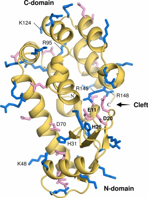
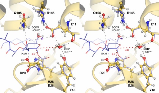
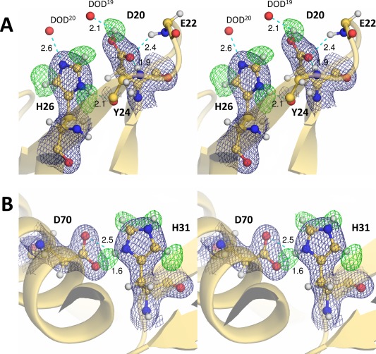
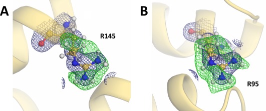
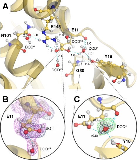
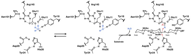
T4 phage lysozyme is an inverting glycoside hydrolase that degrades the murein of bacterial cell walls by cleaving the β-1,4-glycosidic bond. The substitution of the catalytic Thr26 residue to a histidine converts the wild type from an inverting to a retaining enzyme, which implies that the original general acid Glu11 can also act as an acid/base catalyst in the hydrolysis. Here, we have determined the neutron structure of the perdeuterated T26H mutant to clarify the protonation states of Glu11 and the substituted His26, which are key in the retaining reaction. The 2.09-Å resolution structure shows that the imidazole group of His26 is in its singly protonated form in the active site, suggesting that the deprotonated Nɛ2 atom of His26 can attack the anomeric carbon of bound substrate as a nucleophile. The carboxyl group of Glu11 is partially protonated and interacts with the unusual neutral state of the guanidine moiety of Arg145, as well as two heavy water molecules. Considering that one of the water-binding sites has the potential to be occupied by a hydronium ion, the bulk solvent could be the source for the protonation of Glu11. The respective protonation states of Glu11 and His26 are consistent with the bond lengths determined by an unrestrained refinement of the high-resolution X-ray structure of T26H at 1.04-Å resolution. The detail structural information, including the coordinates of the deuterium atoms in the active site, provides insight into the distinctively different catalytic activities of the mutant and wild type enzymes.

摘要

T4噬菌体溶菌酶是一种转化糖苷水解酶,通过切割β-1,4-糖苷键来降解细菌细胞壁的胞壁质。将催化性的苏氨酸26残基替换为组氨酸,可使野生型酶从转化型变为保留型酶,这意味着原来的一般酸谷氨酸11在水解过程中也可作为酸碱催化剂。在此,我们测定了全氘代T26H突变体的中子结构,以阐明谷氨酸11和取代的组氨酸26的质子化状态,这两者在保留反应中起关键作用。分辨率为2.09 Å的结构表明,组氨酸26的咪唑基团在活性位点处于单质子化形式,这表明组氨酸26去质子化的Nɛ2原子可作为亲核试剂攻击结合底物的异头碳。谷氨酸11的羧基部分质子化,并与精氨酸145胍基部分的异常中性状态以及两个重水分子相互作用。考虑到其中一个水结合位点有可能被水合氢离子占据,大量溶剂可能是谷氨酸11质子化的来源。谷氨酸11和组氨酸26各自的质子化状态与分辨率为1.04 Å的T26H高分辨率X射线结构无约束精修所确定的键长一致。详细的结构信息,包括活性位点中氘原子的坐标,为深入了解突变体和野生型酶截然不同的催化活性提供了依据。

https://cdn.ncbi.nlm.nih.gov/pmc/blobs/d9ab/5606550/344704098d07/PRO-26-1953-g006.jpg
https://cdn.ncbi.nlm.nih.gov/pmc/blobs/d9ab/5606550/d8b0c6ce4d6e/PRO-26-1953-g001.jpg
https://cdn.ncbi.nlm.nih.gov/pmc/blobs/d9ab/5606550/8f0fcabd6461/PRO-26-1953-g002.jpg
https://cdn.ncbi.nlm.nih.gov/pmc/blobs/d9ab/5606550/8d52661beca0/PRO-26-1953-g003.jpg
https://cdn.ncbi.nlm.nih.gov/pmc/blobs/d9ab/5606550/1ebcc6f64f3c/PRO-26-1953-g004.jpg
https://cdn.ncbi.nlm.nih.gov/pmc/blobs/d9ab/5606550/91d074ea0543/PRO-26-1953-g005.jpg
https://cdn.ncbi.nlm.nih.gov/pmc/blobs/d9ab/5606550/344704098d07/PRO-26-1953-g006.jpg

相似文献

1
Neutron structure of the T26H mutant of T4 phage lysozyme provides insight into the catalytic activity of the mutant enzyme and how it differs from that of wild type.T4噬菌体溶菌酶T26H突变体的中子结构有助于深入了解突变酶的催化活性及其与野生型酶催化活性的差异。
Protein Sci. 2017 Oct;26(10):1953-1963. doi: 10.1002/pro.3230. Epub 2017 Jul 25.
2
The pK values of the catalytic residues in the retaining glycoside hydrolase T26H mutant of T4 lysozyme.T4 溶菌酶 T26H 突变体中催化残基的 pK 值。
Protein Sci. 2019 Mar;28(3):620-632. doi: 10.1002/pro.3562. Epub 2019 Jan 12.
3
Structural basis of the conversion of T4 lysozyme into a transglycosidase by reengineering the active site.通过对活性位点进行重新设计将T4溶菌酶转化为转糖基酶的结构基础。
Proc Natl Acad Sci U S A. 1999 Aug 3;96(16):8949-54. doi: 10.1073/pnas.96.16.8949.
4
Structure-based design of a lysozyme with altered catalytic activity.具有改变催化活性的溶菌酶的基于结构的设计。
Nat Struct Biol. 1995 Nov;2(11):1007-11. doi: 10.1038/nsb1195-1007.
5
Lysozyme: a model enzyme in protein crystallography.溶菌酶:蛋白质晶体学中的一种模型酶。
EXS. 1996;75:185-222. doi: 10.1007/978-3-0348-9225-4_11.
6
Structural and thermodynamic analysis of the binding of solvent at internal sites in T4 lysozyme.T4溶菌酶内部位点溶剂结合的结构与热力学分析
Protein Sci. 2001 May;10(5):1067-78. doi: 10.1110/ps.02101.
7
Size versus polarizability in protein-ligand interactions: binding of noble gases within engineered cavities in phage T4 lysozyme.蛋白质-配体相互作用中的尺寸与极化率:噬菌体T4溶菌酶工程化空腔内稀有气体的结合
J Mol Biol. 2000 Sep 29;302(4):955-77. doi: 10.1006/jmbi.2000.4063.
8
Modeling protein-small molecule interactions: structure and thermodynamics of noble gases binding in a cavity in mutant phage T4 lysozyme L99A.蛋白质-小分子相互作用建模:突变型噬菌体T4溶菌酶L99A中空腔中稀有气体结合的结构与热力学
J Mol Biol. 2000 Sep 29;302(4):979-89. doi: 10.1006/jmbi.2000.4064.
9
Direct determination of protonation states and visualization of hydrogen bonding in a glycoside hydrolase with neutron crystallography.通过中子晶体学直接测定糖苷水解酶中的质子化状态并可视化氢键。
Proc Natl Acad Sci U S A. 2015 Oct 6;112(40):12384-9. doi: 10.1073/pnas.1504986112. Epub 2015 Sep 21.
10
Structural analysis of a non-contiguous second-site revertant in T4 lysozyme shows that increasing the rigidity of a protein can enhance its stability.T4溶菌酶中一个非连续第二位点回复突变体的结构分析表明,增加蛋白质的刚性可以提高其稳定性。
J Mol Biol. 1999 Oct 8;292(5):1111-20. doi: 10.1006/jmbi.1999.3102.

引用本文的文献

1
Creation of Cross-Linked Crystals With Intermolecular Disulfide Bonds Connecting Symmetry-Related Molecules Allows Retention of Tertiary Structure in Different Solvent Conditions.通过连接对称相关分子的分子间二硫键创建交联晶体,可在不同溶剂条件下保留三级结构。
Front Mol Biosci. 2022 Jun 8;9:908394. doi: 10.3389/fmolb.2022.908394. eCollection 2022.
2
Aspartate or arginine? Validated redox state X-ray structures elucidate mechanistic subtleties of Fe = O formation in bacterial dye-decolorizing peroxidases.天冬氨酸或精氨酸?经证实的氧化还原态 X 射线结构阐明了细菌染料脱色过氧化物酶中 Fe=O 形成的机制细微差别。
J Biol Inorg Chem. 2021 Oct;26(7):743-761. doi: 10.1007/s00775-021-01896-2. Epub 2021 Sep 3.
3

本文引用的文献

1
Processing of X-ray diffraction data collected in oscillation mode.振荡模式下收集的X射线衍射数据的处理。
Methods Enzymol. 1997;276:307-26. doi: 10.1016/S0076-6879(97)76066-X.
2
"Newton's cradle" proton relay with amide-imidic acid tautomerization in inverting cellulase visualized by neutron crystallography.通过中子晶体学可视化的、具有酰胺-亚胺酸互变异构的“牛顿摆”质子传递在转化型纤维素酶中
Sci Adv. 2015 Aug 21;1(7):e1500263. doi: 10.1126/sciadv.1500263. eCollection 2015 Aug.
3
Neutron protein crystallography: A complementary tool for locating hydrogens in proteins.
Serial Femtosecond Zero Dose Crystallography Captures a Water-Free Distal Heme Site in a Dye-Decolorising Peroxidase to Reveal a Catalytic Role for an Arginine in Fe =O Formation.
串联飞秒零剂量晶体学捕获无水分的远位血红素在脱染料过氧化物酶中的位置,揭示了精氨酸在 Fe=O 形成中的催化作用。
Angew Chem Int Ed Engl. 2020 Nov 23;59(48):21656-21662. doi: 10.1002/anie.202008622. Epub 2020 Sep 23.
4
Visualizing the protons in a metalloenzyme electron proton transfer pathway.可视化金属酶电子质子转移途径中的质子。
Proc Natl Acad Sci U S A. 2020 Mar 24;117(12):6484-6490. doi: 10.1073/pnas.1918936117. Epub 2020 Mar 9.
5
The pK values of the catalytic residues in the retaining glycoside hydrolase T26H mutant of T4 lysozyme.T4 溶菌酶 T26H 突变体中催化残基的 pK 值。
Protein Sci. 2019 Mar;28(3):620-632. doi: 10.1002/pro.3562. Epub 2019 Jan 12.
中子蛋白质晶体学:一种用于确定蛋白质中氢原子位置的互补工具。
Arch Biochem Biophys. 2016 Jul 15;602:48-60. doi: 10.1016/j.abb.2015.11.033. Epub 2015 Nov 22.
4
Direct determination of protonation states and visualization of hydrogen bonding in a glycoside hydrolase with neutron crystallography.通过中子晶体学直接测定糖苷水解酶中的质子化状态并可视化氢键。
Proc Natl Acad Sci U S A. 2015 Oct 6;112(40):12384-9. doi: 10.1073/pnas.1504986112. Epub 2015 Sep 21.
5
New insights into the enzymatic mechanism of human chitotriosidase (CHIT1) catalytic domain by atomic resolution X-ray diffraction and hybrid QM/MM.通过原子分辨率X射线衍射和量子力学/分子力学混合方法对人几丁质酶(CHIT1)催化结构域酶促机制的新见解。
Acta Crystallogr D Biol Crystallogr. 2015 Jul;71(Pt 7):1455-70. doi: 10.1107/S139900471500783X. Epub 2015 Jun 30.
6
Protonation and geometry of histidine rings.组氨酸环的质子化与几何结构。
Acta Crystallogr D Biol Crystallogr. 2015 Jul;71(Pt 7):1444-54. doi: 10.1107/S1399004715007816. Epub 2015 Jun 30.
7
Arginine: Its pKa value revisited.精氨酸:重新审视其pKa值。
Protein Sci. 2015 May;24(5):752-61. doi: 10.1002/pro.2647. Epub 2015 Mar 22.
8
The IMAGINE instrument: first neutron protein structure and new capabilities for neutron macromolecular crystallography.IMAGINE仪器:首个中子蛋白质结构及中子大分子晶体学的新功能
Acta Crystallogr D Biol Crystallogr. 2013 Oct;69(Pt 10):2157-60. doi: 10.1107/S0907444913019604. Epub 2013 Sep 20.
9
Near-atomic resolution neutron crystallography on perdeuterated Pyrococcus furiosus rubredoxin: implication of hydronium ions and protonation state equilibria in redox changes.嗜热栖热菌铁氧化还原蛋白全氘代物的近原子分辨率中子晶体学:水合氢离子和质子化状态平衡在氧化还原变化中的作用
Angew Chem Int Ed Engl. 2013 Jan 14;52(3):1022-5. doi: 10.1002/anie.201207071. Epub 2012 Dec 6.
10
Determination of the rotational dynamics and pH dependence of the hydrogen exchange rates of the arginine guanidino group using NMR spectroscopy.利用 NMR 光谱法测定精氨酸胍基的旋转动力学和 pH 值依赖性的氢交换速率。
J Biomol NMR. 1995 Jul;6(1):59-66. doi: 10.1007/BF00417492.