Luo Zhenli, Xu Wenhuan, Ma Sai, Qiao Hongyu, Gao Lei, Zhang Ran, Yang Bo, Qiu Ya, Chen Jiangwei, Zhang Ming, Tao Bo, Cao Feng, Wang Yabin
Department of Cardiology, Xijing Hospital, Fourth Military Medical University, Xi'an 710032, China.
Training and Postgraduate Management Department, Medical Administrative Division, Chinese PLA General Hospital, Beijing 100853, China.
Oxid Med Cell Longev. 2017;2017:3018190. doi: 10.1155/2017/3018190. Epub 2017 Jun 21.
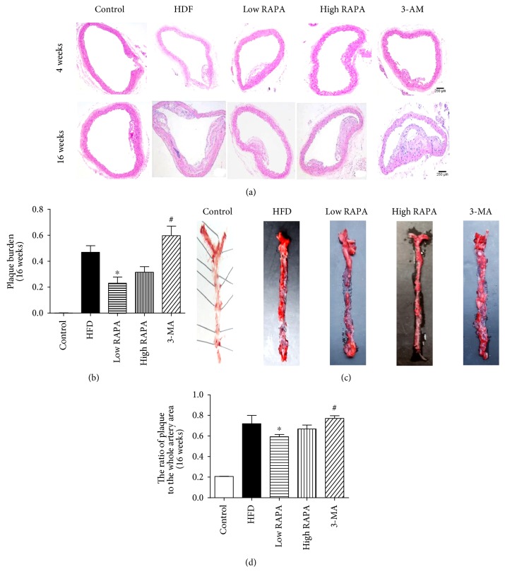
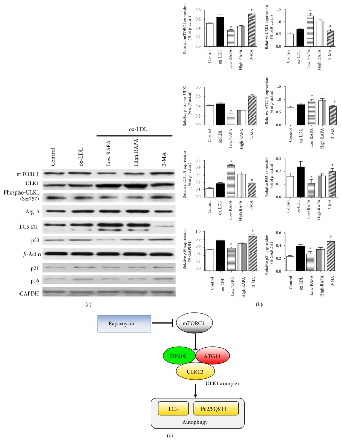
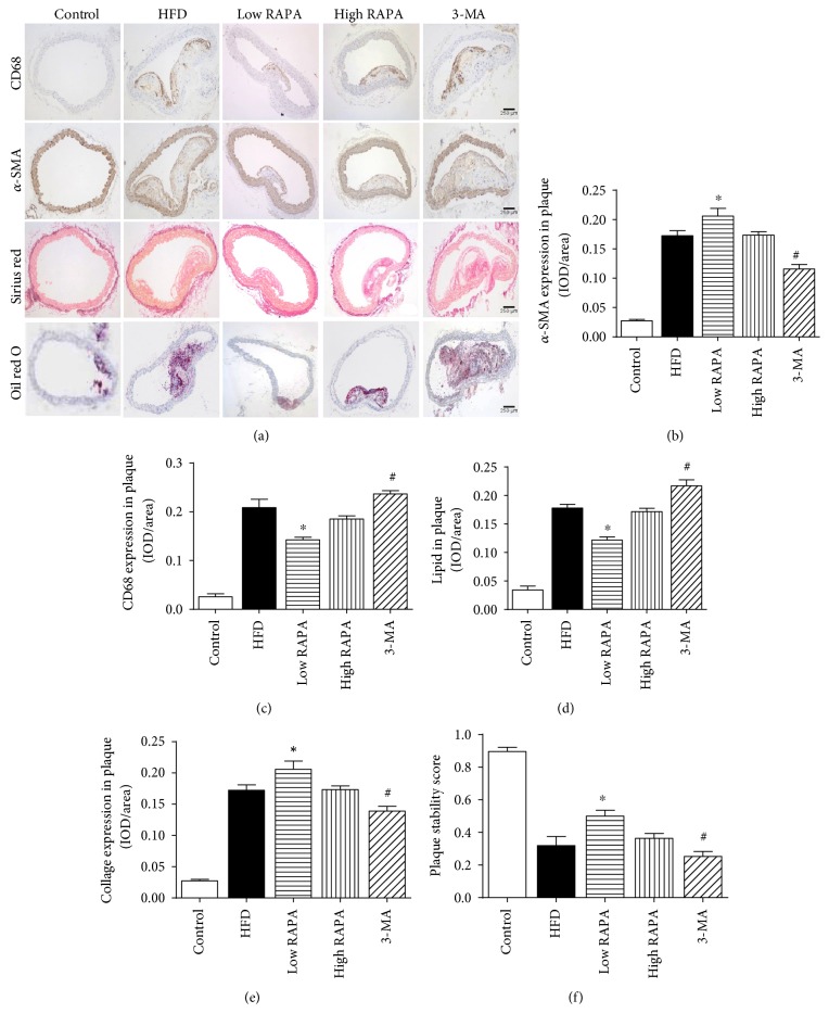
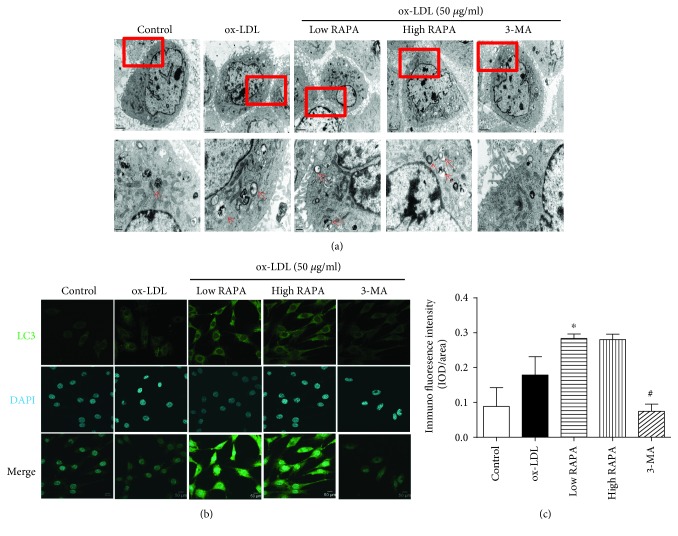
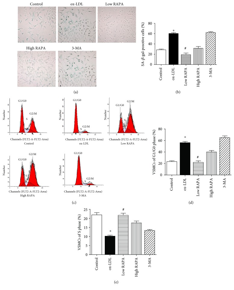
In order to investigate the effects of autophagy induced by rapamycin in the development of atherosclerosis plaque we established murine atherosclerosis model which was induced in ApoE mice by high fat and cholesterol diet (HFD) for 16 weeks. Rapamycin and 3-Methyladenine (MA) were used as autophagy inducer and inhibitor respectively. The plaque areas in aortic artery were detected with HE and Oil Red O staining. Immunohistochemical staining were applied to investigate content of plaque respectively. In contrast to control and 3-MA groups, rapamycin could inhibit atherosclerosis progression. Rapamycin was able to increase collagen content and a-SMA distribution relatively, as well as decrease necrotic core area. Then we used MOVAS and culture with ox-LDL for 72 h to induce smooth muscle-derived foam cell model in vitro. Rapamycin and 3-MA were cultured together respectively. Flow cytometry assay and SA--Gal staining experiments were performed to detect survival and senescence of VSMCs. Western blot analysis were utilized to analyze the levels of protein expression. We found that rapamycin could promote ox-LDL-induced VSMCs autophagy survival and alleviate cellular senescence, in comparison to control and 3-MA groups. Western blot analysis showed that rapamycin could upregulate ULK1, ATG13 and downregulate mTORC1 and p53 protein expression.
为了研究雷帕霉素诱导的自噬在动脉粥样硬化斑块形成中的作用,我们建立了小鼠动脉粥样硬化模型,该模型通过高脂高胆固醇饮食(HFD)诱导ApoE小鼠16周。雷帕霉素和3-甲基腺嘌呤(MA)分别用作自噬诱导剂和抑制剂。用苏木精-伊红(HE)和油红O染色检测主动脉中的斑块面积。应用免疫组织化学染色分别研究斑块的成分。与对照组和3-MA组相比,雷帕霉素可抑制动脉粥样硬化进展。雷帕霉素能够相对增加胶原蛋白含量和α-平滑肌肌动蛋白(α-SMA)分布,同时减少坏死核心面积。然后我们使用小鼠主动脉血管平滑肌细胞(MOVAS)并在氧化型低密度脂蛋白(ox-LDL)中培养72小时,以在体外诱导平滑肌来源的泡沫细胞模型。雷帕霉素和3-MA分别共同培养。进行流式细胞术检测和衰老相关β-半乳糖苷酶(SA-β-Gal)染色实验,以检测血管平滑肌细胞(VSMCs)的存活和衰老情况。利用蛋白质免疫印迹分析来分析蛋白质表达水平。我们发现,与对照组和3-MA组相比,雷帕霉素可促进ox-LDL诱导的VSMCs自噬存活并减轻细胞衰老。蛋白质免疫印迹分析表明,雷帕霉素可上调Unc-51样激酶1(ULK1)、自噬相关蛋白13(ATG13),并下调哺乳动物雷帕霉素靶蛋白复合物1(mTORC1)和p53蛋白表达。