Martínez-Rivera Freddyson J, Pérez-Laspiur Juliana, Santiago-Gascot María E, Alemán-Reyes Abner G, García-Santiago Emanuel, Rodríguez-Pérez Yolanda, Calo-Guadalupe Cristhian, Otero-Pagán Inelia, Ayala-Pagán Roxsana N, Martínez Magdiel, Cantres-Rosario Yisel M, Meléndez Loyda M, Barreto-Estrada Jennifer L
Department of Anatomy and Neurobiology, Medical Sciences Campus, University of Puerto Rico, San Juan, Puerto Rico, United States of America.
Translational Proteomics Center-RCMI, Medical Sciences Campus, University of Puerto Rico, San Juan, Puerto Rico, United States of America.
PLoS One. 2017 Jul 18;12(7):e0180409. doi: 10.1371/journal.pone.0180409. eCollection 2017.
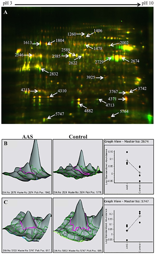
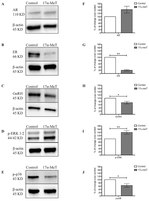
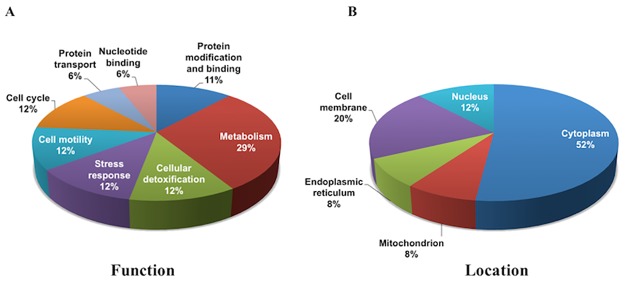
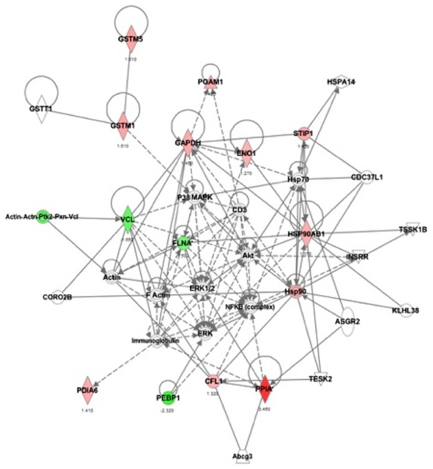
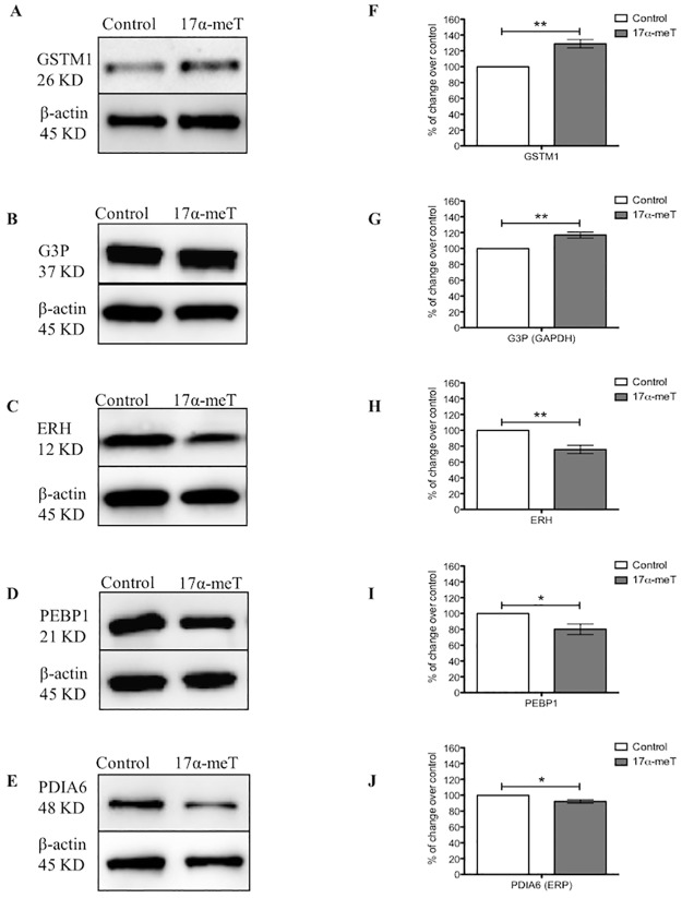
The abuse of anabolic androgenic steroids (AAS) has been considered a major public health problem during decades. Supraphysiological doses of AAS may lead to a variety of neuroendocrine problems. Precisely, the hypothalamic-pituitary-gonadal (HPG) axis is one of the body systems that is mainly influenced by steroidal hormones. Fluctuations of the hormonal milieu result in alterations of reproductive function, which are made through changes in hypothalamic neurons expressing gonadotropin-releasing hormone (GnRH). In fact, previous studies have shown that AAS modulate the activity of these neurons through steroid-sensitive afferents. To increase knowledge about the cellular mechanisms induced by AAS in GnRH neurons, we performed proteomic analyses of the murine hypothalamic GT1-7 cell line after exposure to 17α-methyltestosterone (17α-meT; 1 μM). These cells represent a good model for studying regulatory processes because they exhibit the typical characteristics of GnRH neurons, and respond to compounds that modulate GnRH in vivo. Two-dimensional difference in gel electrophoresis (2D-DIGE) and mass spectrometry analyses identified a total of 17 different proteins that were significantly affected by supraphysiological levels of AAS. Furthermore, pathway analyses showed that modulated proteins were mainly associated to glucose metabolism, drug detoxification, stress response and cell cycle. Validation of many of these proteins, such as GSTM1, ERH, GAPDH, PEBP1 and PDIA6, were confirmed by western blotting. We further demonstrated that AAS exposure decreased expression of estrogen receptors and GnRH, while two important signaling pathway proteins p-ERK, and p-p38, were modulated. Our results suggest that steroids have the capacity to directly affect the neuroendocrine system by modulating key cellular processes for the control of reproductive function.
几十年来,合成代谢雄激素类固醇(AAS)的滥用一直被视为一个重大的公共卫生问题。超生理剂量的AAS可能会导致各种神经内分泌问题。确切地说,下丘脑-垂体-性腺(HPG)轴是主要受甾体激素影响的身体系统之一。激素环境的波动会导致生殖功能的改变,这是通过表达促性腺激素释放激素(GnRH)的下丘脑神经元的变化实现的。事实上,先前的研究表明,AAS通过类固醇敏感传入神经调节这些神经元的活性。为了增加对AAS在GnRH神经元中诱导的细胞机制的了解,我们对暴露于17α-甲基睾酮(17α-meT;1μM)后的小鼠下丘脑GT1-7细胞系进行了蛋白质组学分析。这些细胞代表了一个研究调节过程的良好模型,因为它们表现出GnRH神经元的典型特征,并对体内调节GnRH的化合物有反应。二维差异凝胶电泳(2D-DIGE)和质谱分析共鉴定出17种受超生理水平AAS显著影响的不同蛋白质。此外,通路分析表明,受调节的蛋白质主要与葡萄糖代谢、药物解毒、应激反应和细胞周期有关。许多这些蛋白质,如谷胱甘肽S-转移酶M1(GSTM1)、富含脯氨酸的蛋白质(ERH)、甘油醛-3-磷酸脱氢酶(GAPDH)、磷脂酰乙醇胺结合蛋白1(PEBP1)和蛋白二硫键异构酶A6(PDIA6),通过蛋白质印迹法得到了验证。我们进一步证明,AAS暴露会降低雌激素受体和GnRH的表达,同时两种重要的信号通路蛋白磷酸化细胞外信号调节激酶(p-ERK)和磷酸化p38(p-p38)也受到了调节。我们的结果表明,类固醇有能力通过调节控制生殖功能的关键细胞过程来直接影响神经内分泌系统。