Inje University College of Medicine Haeundae-gu Busan Korea.
Brain Behav. 2017 May 23;7(7):e00730. doi: 10.1002/brb3.730. eCollection 2017 Jul.
This study aimed to investigate the differences in brain morphology according to handedness.
Forty-two healthy subjects were enrolled (21 right-handers and 21 nonright-handers). The two groups were classified according to the Edinburgh Handedness Inventory. Measures of cortical morphology, such as thickness, surface area, volume, and curvature, and the volumes of subcortical structures, such as the amygdala, caudate, hippocampus, globus pallidus, putamen, and thalamus, were compared between the groups according to handedness using whole-brain 3D T1-weighted MRI. In addition, we investigated the white matter differences between the groups using diffusion tensor imaging. Moreover, we quantified correlations between the handedness scales of the Edinburgh Handedness Inventory and each measure of different brain morphologies.
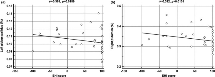
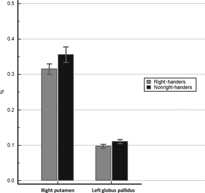
The volumes of the right putamen and left globus pallidus in nonright-handed participants were significantly larger than those who were right-handed (0.3559 vs. 0.3155%, =.0028; 0.1101 vs. 0.0975%, =.0025; respectively). Moreover, the volumes of the right putamen and left globus pallidus were negatively correlated with the handedness scales of the Edinburgh Handedness Inventory (= -.392, =.0101; = -.361, =.0189; respectively). However, the cortex morphology and the other subcortical volumes were not significantly different between the two groups. In addition, we did not find any white matter differences between the groups.
We demonstrated that there were significant differences in brain morphology between right-handers and nonright-handers, especially in the basal ganglia, which could produce differences in motor control according to handedness.
本研究旨在探讨大脑形态根据利手的差异。
共纳入 42 名健康受试者(21 名右利手和 21 名非右利手)。两组根据爱丁堡手性量表进行分类。使用全脑 3D T1 加权 MRI 根据利手比较两组皮质形态(如厚度、表面积、体积和曲率)和皮质下结构(如杏仁核、尾状核、海马体、苍白球、壳核和丘脑)的体积差异。此外,我们使用弥散张量成像研究了两组之间的白质差异。还量化了爱丁堡手性量表的利手评分与不同脑形态的每个测量值之间的相关性。
非右利手参与者的右侧壳核和左侧苍白球体积明显大于右利手者(0.3559 对 0.3155%,= 0.0028;0.1101 对 0.0975%,= 0.0025;分别)。此外,右侧壳核和左侧苍白球体积与爱丁堡手性量表的利手评分呈负相关(= -0.392,= 0.0101;= -0.361,= 0.0189;分别)。然而,两组皮质形态和其他皮质下体积无明显差异。此外,我们未发现两组之间存在任何白质差异。
我们表明,右利手和非右利手之间的大脑形态存在显著差异,尤其是在基底节,这可能根据利手产生运动控制的差异。