Department of Radiology Eye and ENT Hospital of Shanghai Medical School Fudan University Shanghai China.
Department of Radiotherapy Eye and ENT Hospital of Shanghai Medical School Fudan University Shanghai China.
Brain Behav. 2017 May 22;7(7):e00731. doi: 10.1002/brb3.731. eCollection 2017 Jul.
Our study aimed to explore the feasibility of manganese-enhanced magnetic resonance imaging (MEMRI) combined with visual evoked potentials (VEP) and auditory evoked visual cortex responses (AVR) in evaluating for the establishment of visual/auditory compensatory pathways after monocular blindness.
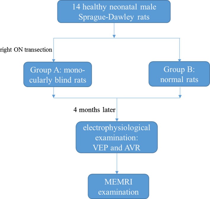
A total of 14 healthy neonatal male Sprague-Dawley rats were randomly divided into two groups (= 7 for Groups A and B). Right optic nerve (ON) transection was performed on the 7 rats of Group A to obtain a monocularly blind model, and the 7 rats of Group B were chosen as the control group. Four months later, 400 mmol MnCl was injected into the left eye in both groups via intravitreal injection. The changes in the visual pathways projected from the blind eye and the remaining eye in Group A and the normal eyes in Group B were compared to determine if new visual compensatory pathways were established. Additionally, VEP tests were performed to determine complete blindness, and AVR examinations were performed to help identify the generation of auditory compensatory function.
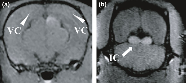
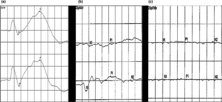
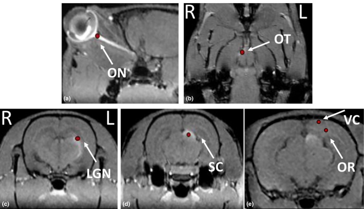
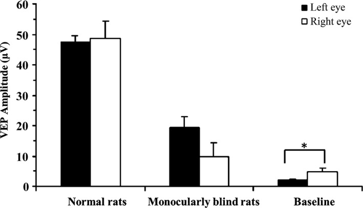
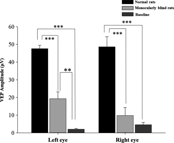
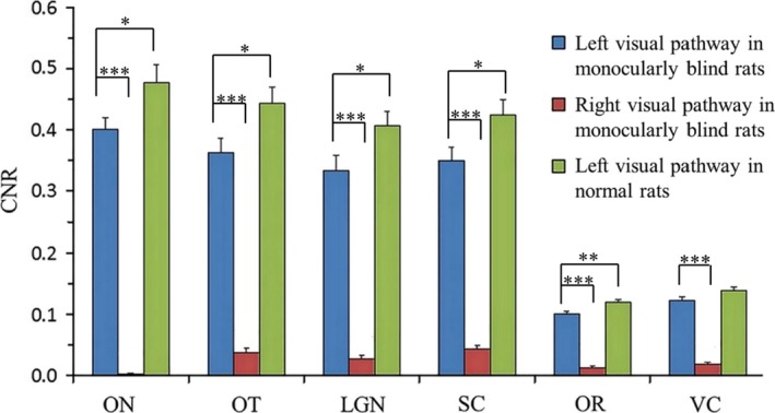
The VEP test indicated complete visual loss after ON transection. In the monocularly blind rats, the contrast-to-noise ratio (CNR) of ON, optic tract (OT), lateral geniculate nucleus (LGN), superior colliculus (SC), optic radiation (OR) and visual cortex (VC) of visual pathway projected from the left eye was significantly higher than that of the right pathway (<.001). Moreover, the CNR of ON, OT, LGN, SC, OR and VC in the visual pathway projected from the left eye of monocularly blind rats was significantly lower than those of normal rats (<.05). The AVR results revealed that the corresponding bilateral visual cortex in monocularly blind rats did not respond to the auditory stimulus or showed dissimilation with the low frequency.
MEMRI combined with electrophysiology, including VEP and AVR, may be potentially helpful in the evaluation of the possible generation of new visual/auditory compensatory pathways after monocular blindness.
本研究旨在探讨锰增强磁共振成像(MEMRI)结合视觉诱发电位(VEP)和听觉诱发电经视觉皮层反应(AVR)评估单侧盲后视觉/听觉补偿通路建立的可行性。
将 14 只健康新生雄性 Sprague-Dawley 大鼠随机分为两组(A 组和 B 组各 7 只)。A 组 7 只大鼠右眼视神经(ON)切断,建立单侧盲模型,B 组 7 只大鼠作为对照组。4 个月后,两组大鼠均经玻璃体腔注射 400mmol MnCl2。比较 A 组盲眼和保留眼以及 B 组正常眼投射的视觉通路的变化,以确定是否建立新的视觉补偿通路。此外,进行 VEP 测试以确定完全失明,并进行 AVR 检查以帮助识别听觉补偿功能的产生。
ON 切断后,VEP 测试表明完全丧失视觉。在单侧盲大鼠中,左眼投射的视路中 ON、视束(OT)、外侧膝状体核(LGN)、上丘(SC)、视辐射(OR)和视觉皮层(VC)的对比噪声比(CNR)明显高于右眼(<0.001)。此外,单侧盲大鼠左眼投射视路中 ON、OT、LGN、SC、OR 和 VC 的 CNR 明显低于正常大鼠(<0.05)。AVR 结果显示,单侧盲大鼠双侧相应视皮层对听觉刺激无反应或低频出现异化。
MEMRI 结合电生理学,包括 VEP 和 AVR,可能有助于评估单侧盲后新的视觉/听觉补偿通路的产生。