Wei Yantao, Jiang Hong, Shi Yingying, Qu Dongyi, Gregori Giovanni, Zheng Fang, Rundek Tatjana, Wang Jianhua
Zhongshan Ophthalmic Center, Sun Yat-sen University, Guangzhou, Guangdong, China
Bascom Palmer Eye Institute, University of Miami Miller School of Medicine, Miami, Florida, United States
Invest Ophthalmol Vis Sci. 2017 Jul 1;58(9):3804-3817. doi: 10.1167/iovs.17-21460.
To characterize age-related alterations in the retinal microcirculation, microvascular network, and microstructure in healthy subjects.
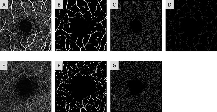
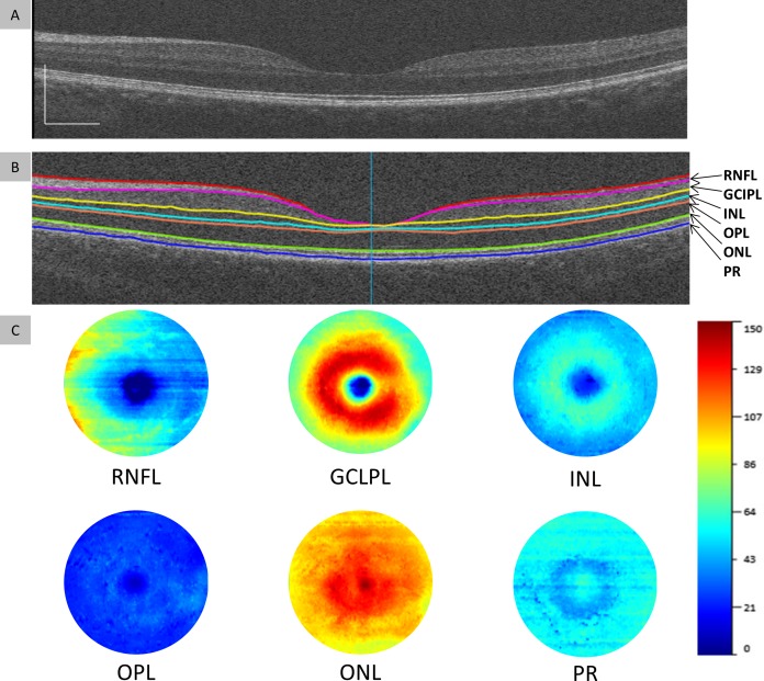
Seventy-four healthy subjects aged from 18 to 82 years were recruited and divided into four age groups (G1 with age <35 years, G2 with age 35 ∼ 49 years, G3 with age 50 ∼ 64 years, and G4 with age ≥65 years). Custom ultra-high resolution optical coherence tomography (UHR-OCT) was used to acquire six intraretinal layers of the macula. OCT angiography (OCTA) was used to image the retinal microvascular network. The retinal blood flow velocity (BFV) was measured using a Retinal Function Imager (RFI).
Compared to G1, G2 had significant thinning of the retinal nerve fiber layer (RNFL) (P < 0.05), while G3 had thinning of the RNFL and ganglion cell and inner plexiform layer (GCIPL) (P < 0.05), in addition to thickening of the outer plexiform layer (OPL) and photoreceptor layer (PR) (P < 0.05). G4 had loss in retinal vessel density, thinning in RNFL and GCIPL, and decrease in venular BFV, in addition to thickening of the OPL and PR (P < 0.05). Age was negatively related to retinal vessel densities, the inner retinal layers, and venular BFV (P < 0.05). By contrast, age was positively related to OPL and PR (P < 0.05).
During aging, decreases in retinal vessel density, inner retinal layer thickness, and venular BFV were evident and impacted each other as observed by simultaneous changes in multiple retinal components.
描述健康受试者视网膜微循环、微血管网络及微观结构的年龄相关性变化。
招募了74名年龄在18至82岁之间的健康受试者,并将其分为四个年龄组(G1组年龄<35岁,G2组年龄35至49岁,G3组年龄50至64岁,G4组年龄≥65岁)。使用定制的超高分辨率光学相干断层扫描(UHR-OCT)获取黄斑的六层视网膜内结构。采用光学相干断层扫描血管造影(OCTA)对视网膜微血管网络进行成像。使用视网膜功能成像仪(RFI)测量视网膜血流速度(BFV)。
与G1组相比,G2组视网膜神经纤维层(RNFL)显著变薄(P<0.05),而G3组除了外丛状层(OPL)和光感受器层(PR)增厚(P<0.05)外,RNFL以及神经节细胞和内丛状层(GCIPL)均变薄(P<0.05)。G4组除了OPL和PR增厚(P<0.05)外,视网膜血管密度降低,RNFL和GCIPL变薄,小静脉BFV下降(P<0.05)。年龄与视网膜血管密度、视网膜内层及小静脉BFV呈负相关(P<0.05)。相比之下,年龄与OPL和PR呈正相关(P<0.05)。
在衰老过程中,视网膜血管密度、视网膜内层厚度和小静脉BFV的降低是明显的,并且通过多个视网膜成分的同时变化观察到它们之间相互影响。