• 文献检索
  • 文档翻译
  • 深度研究
  • 学术资讯
  • Suppr Zotero 插件Zotero 插件
  • 邀请有礼
  • 套餐&价格
  • 历史记录
应用&插件
Suppr Zotero 插件Zotero 插件浏览器插件Mac 客户端Windows 客户端微信小程序
定价
高级版会员购买积分包购买API积分包
服务
文献检索文档翻译深度研究API 文档MCP 服务
关于我们
关于 Suppr公司介绍联系我们用户协议隐私条款
关注我们

Suppr 超能文献

核心技术专利:CN118964589B侵权必究
粤ICP备2023148730 号-1Suppr @ 2026

文献检索

告别复杂PubMed语法,用中文像聊天一样搜索,搜遍4000万医学文献。AI智能推荐,让科研检索更轻松。

立即免费搜索

文件翻译

保留排版,准确专业,支持PDF/Word/PPT等文件格式,支持 12+语言互译。

免费翻译文档

深度研究

AI帮你快速写综述,25分钟生成高质量综述,智能提取关键信息,辅助科研写作。

立即免费体验

使用基于光学相干断层扫描的微血管造影术评估青光眼患者视乳头周围视网膜神经纤维层的血管微循环

Peripapillary Retinal Nerve Fiber Layer Vascular Microcirculation in Glaucoma Using Optical Coherence Tomography-Based Microangiography.

作者信息

Chen Chieh-Li, Zhang Anqi, Bojikian Karine D, Wen Joanne C, Zhang Qinqin, Xin Chen, Mudumbai Raghu C, Johnstone Murray A, Chen Philip P, Wang Ruikang K

机构信息

Department of Bioengineering, University of Washington, Seattle, Washington, United States 2Department of Ophthalmology, University of Washington, Seattle, Washington, United States.

Department of Bioengineering, University of Washington, Seattle, Washington, United States.

出版信息

Invest Ophthalmol Vis Sci. 2016 Jul 1;57(9):OCT475-85. doi: 10.1167/iovs.15-18909.

DOI:10.1167/iovs.15-18909
PMID:27442341
原文链接:https://pmc.ncbi.nlm.nih.gov/articles/PMC4968914/
Abstract

PURPOSE

To investigate the vascular microcirculation changes in the retinal nerve fiber layer (RNFL) in normal, glaucoma suspect, and open-angle glaucoma (OAG) groups using optical coherence tomography-based microangiography (OMAG).

METHODS

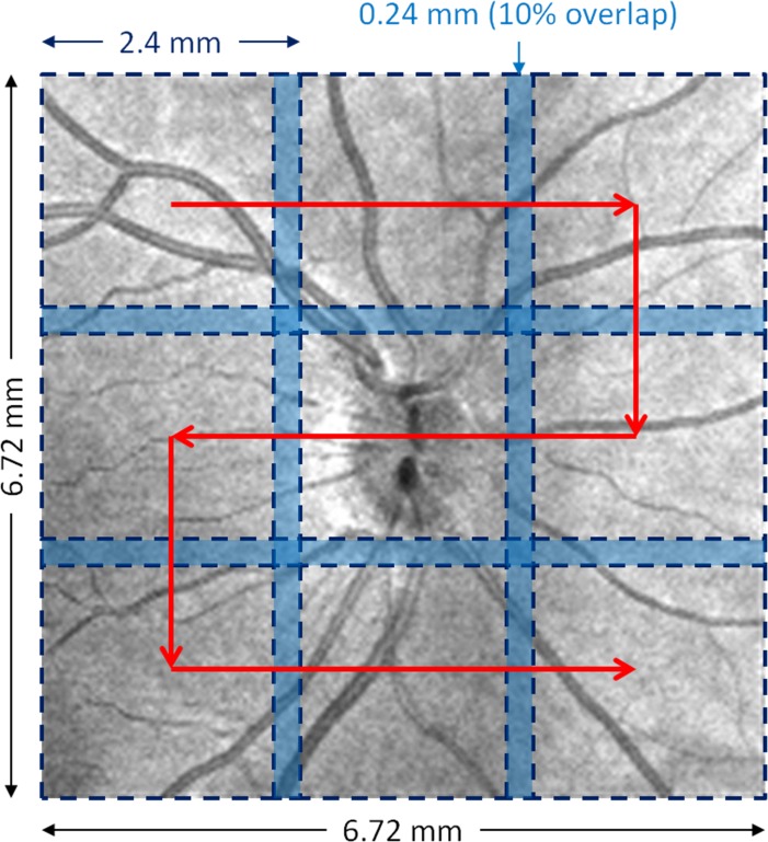
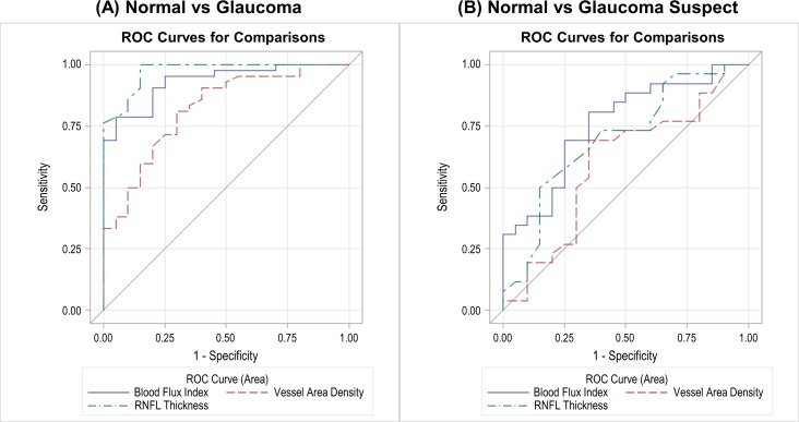
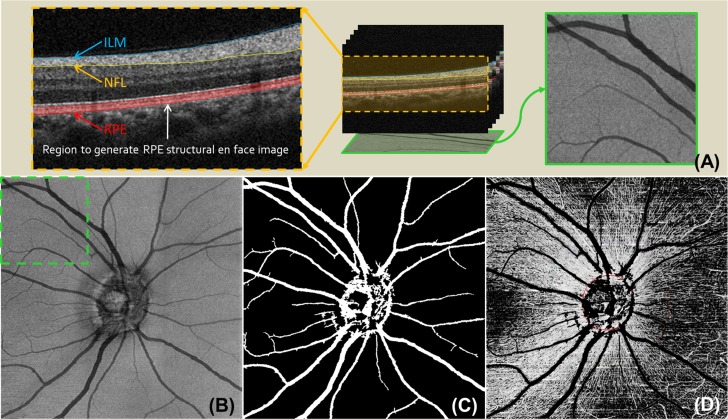
One eye from each subject was scanned with a Cirrus HD-OCT 5000-based OMAG prototype system montage scanning protocol centered at the optic nerve head (ONH). Blood flow signals were extracted using OMAG algorithm. Retinal nerve fiber layer vascular microcirculation was measured by calculating the blood flux index and vessel area density within a 1.2-mm width annulus centered at the ONH with exclusion of big retinal vessels. One-way ANOVA were performed to analyze the RNFL microcirculation among groups. Linear-regression models were constructed to analyze the correlation between RNFL microcirculation and clinical parameters. Discrimination capabilities of the flow metrics were assessed with the area under the receiver operating characteristic curve (AROC).

RESULTS

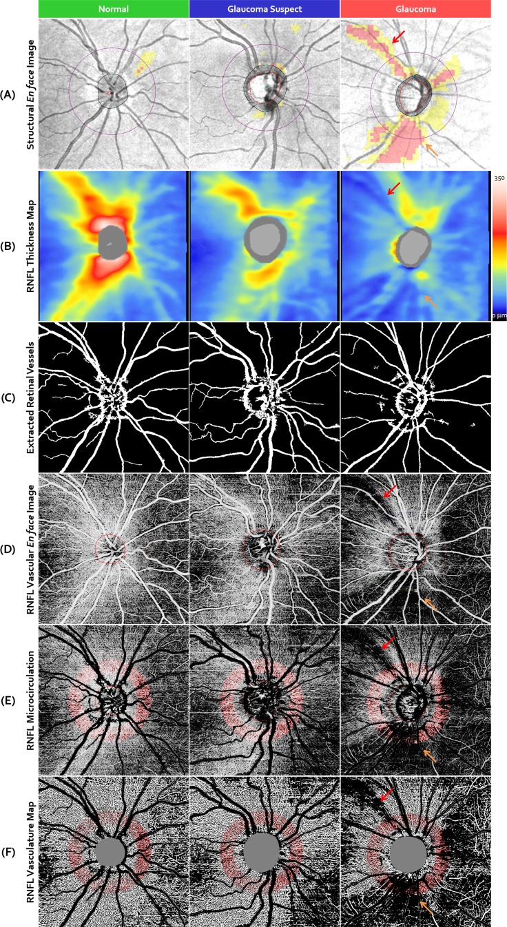
Twenty normal, 26 glaucoma suspect, and 42 OAG subjects were enrolled. Eyes from OAG subjects and glaucoma suspects showed significantly lower blood flux index compared with normal eyes (P ≤ 0.0015). Retinal nerve fiber layer blood flow metrics showed significant correlations with visual field indices and structural changes in glaucomatous eyes (P ≤ 0.0123). Similar discrimination capability of blood flux index compared with RNFL thickness was found in both disease groups.

CONCLUSIONS

Peripapillary RNFL vascular microcirculation measured as blood flux index by OMAG showed significant differences among OAG, glaucoma suspect, and normal controls and was significantly correlated with functional and structural defects. Retinal nerve fiber layer microcirculation measurement using OMAG may help physicians monitor glaucoma.

摘要

目的

使用基于光学相干断层扫描的微血管造影(OMAG)技术,研究正常组、青光眼可疑组和开角型青光眼(OAG)组视网膜神经纤维层(RNFL)的血管微循环变化。

方法

使用基于Cirrus HD-OCT 5000的OMAG原型系统,以视神经乳头(ONH)为中心的蒙太奇扫描协议对每个受试者的一只眼睛进行扫描。使用OMAG算法提取血流信号。通过计算以ONH为中心、宽度为1.2毫米的环形区域内(排除视网膜大血管)的血流指数和血管面积密度,来测量视网膜神经纤维层的血管微循环。采用单因素方差分析来分析各组之间的RNFL微循环情况。构建线性回归模型来分析RNFL微循环与临床参数之间的相关性。使用受试者操作特征曲线下面积(AROC)评估血流指标的鉴别能力。

结果

纳入20名正常受试者、26名青光眼可疑受试者和42名OAG受试者。与正常眼睛相比,OAG受试者和青光眼可疑受试者的眼睛血流指数显著降低(P≤0.0015)。在青光眼患者眼中,视网膜神经纤维层血流指标与视野指标和结构变化显著相关(P≤0.0123)。在两个疾病组中,发现血流指数与RNFL厚度具有相似的鉴别能力。

结论

通过OMAG测量的视乳头周围RNFL血管微循环血流指数在OAG组、青光眼可疑组和正常对照组之间存在显著差异,并且与功能和结构缺陷显著相关。使用OMAG测量视网膜神经纤维层微循环可能有助于医生监测青光眼。

https://cdn.ncbi.nlm.nih.gov/pmc/blobs/1de8/4968914/31c53c19a083/i1552-5783-57-9-OCT475-f04.jpg
https://cdn.ncbi.nlm.nih.gov/pmc/blobs/1de8/4968914/feb8c90ef50f/i1552-5783-57-9-OCT475-f01.jpg
https://cdn.ncbi.nlm.nih.gov/pmc/blobs/1de8/4968914/2a46b9ba9176/i1552-5783-57-9-OCT475-f02.jpg
https://cdn.ncbi.nlm.nih.gov/pmc/blobs/1de8/4968914/f93a53222f2a/i1552-5783-57-9-OCT475-f03.jpg
https://cdn.ncbi.nlm.nih.gov/pmc/blobs/1de8/4968914/31c53c19a083/i1552-5783-57-9-OCT475-f04.jpg

相似文献

1
Peripapillary Retinal Nerve Fiber Layer Vascular Microcirculation in Glaucoma Using Optical Coherence Tomography-Based Microangiography.使用基于光学相干断层扫描的微血管造影术评估青光眼患者视乳头周围视网膜神经纤维层的血管微循环
Invest Ophthalmol Vis Sci. 2016 Jul 1;57(9):OCT475-85. doi: 10.1167/iovs.15-18909.
2
Peripapillary Retinal Nerve Fiber Layer Vascular Microcirculation in Eyes With Glaucoma and Single-Hemifield Visual Field Loss.青光眼伴单眼半视野缺损患者的视乳头周围视网膜神经纤维层血管微循环
JAMA Ophthalmol. 2017 May 1;135(5):461-468. doi: 10.1001/jamaophthalmol.2017.0261.
3
Glaucoma Diagnostic Ability of the Optical Coherence Tomography Angiography Vessel Density Parameters.光学相干断层扫描血管造影血管密度参数的青光眼诊断能力
Curr Eye Res. 2017 Nov;42(11):1458-1467. doi: 10.1080/02713683.2017.1337157. Epub 2017 Sep 14.
4
Peripapillary and Macular Microcirculation in Glaucoma Patients of African and European Descent Using Optical Coherence Tomography Angiography.用光学相干断层扫描血管造影术研究非洲裔和欧洲裔青光眼患者的视盘周围和黄斑微循环。
J Glaucoma. 2020 Oct;29(10):885-889. doi: 10.1097/IJG.0000000000001629.
5
Optical Coherence Tomography Angiography Vessel Density in Healthy, Glaucoma Suspect, and Glaucoma Eyes.健康眼、青光眼可疑眼和青光眼眼中光学相干断层扫描血管造影的血管密度
Invest Ophthalmol Vis Sci. 2016 Jul 1;57(9):OCT451-9. doi: 10.1167/iovs.15-18944.
6
Parafoveal and optic disc vessel density in patients with obstructive sleep apnea syndrome: an optical coherence tomography angiography study.阻塞性睡眠呼吸暂停综合征患者的旁中心凹和视盘血管密度:一项光学相干断层扫描血管造影研究
Graefes Arch Clin Exp Ophthalmol. 2018 Jul;256(7):1235-1243. doi: 10.1007/s00417-018-3943-7. Epub 2018 Mar 25.
7
Macular Vascular Microcirculation in Eyes With Open-angle Glaucoma Using Different Visual Field Severity Classification Systems.使用不同的视野严重程度分类系统评估开角型青光眼患者的黄斑血管微循环。
J Glaucoma. 2019 Sep;28(9):790-796. doi: 10.1097/IJG.0000000000001308.
8
OCTA vessel density changes in the macular zone in glaucomatous eyes.青光眼患者眼部黄斑区光学相干断层扫描血管密度的变化
Graefes Arch Clin Exp Ophthalmol. 2018 Aug;256(8):1499-1508. doi: 10.1007/s00417-018-3965-1. Epub 2018 Apr 10.
9
Application of optical coherence tomography in glaucoma suspect eyes.光学相干断层扫描在青光眼可疑眼中的应用。
Clin Exp Optom. 2012 Jan;95(1):78-88. doi: 10.1111/j.1444-0938.2011.00654.x. Epub 2011 Oct 10.
10
Quantitative Optical Coherence Tomography Angiography of Radial Peripapillary Capillaries in Glaucoma, Glaucoma Suspect, and Normal Eyes.青光眼、疑似青光眼和正常眼的视盘周围放射状毛细血管的定量光学相干断层扫描血管造影
Am J Ophthalmol. 2016 Oct;170:41-49. doi: 10.1016/j.ajo.2016.07.015. Epub 2016 Jul 25.

引用本文的文献

1
Assessment of Retinal Microcirculation in Primary Open-Angle Glaucoma Using Adaptive Optics and OCT Angiography: Correlation with Structural and Functional Damage.使用自适应光学和光学相干断层扫描血管造影术评估原发性开角型青光眼的视网膜微循环:与结构和功能损害的相关性
J Clin Med. 2025 Jul 14;14(14):4978. doi: 10.3390/jcm14144978.
2
Diabetes-Induced Dysregulation of Peripapillary and Macular Neurovascular Units.糖尿病引起的视乳头周围和黄斑神经血管单元的调节异常。
Invest Ophthalmol Vis Sci. 2025 Jul 1;66(9):10. doi: 10.1167/iovs.66.9.10.
3
Visible-light optical coherence tomography and its applications.

本文引用的文献

1
Optic Disc Perfusion in Primary Open Angle and Normal Tension Glaucoma Eyes Using Optical Coherence Tomography-Based Microangiography.使用基于光学相干断层扫描的微血管造影术评估原发性开角型青光眼和正常眼压性青光眼患者视盘的血流灌注情况
PLoS One. 2016 May 5;11(5):e0154691. doi: 10.1371/journal.pone.0154691. eCollection 2016.
2
Evaluating the effect of pupil dilation on spectral-domain optical coherence tomography measurements and their quality score.评估瞳孔散大对频域光学相干断层扫描测量结果及其质量评分的影响。
BMC Ophthalmol. 2015 Dec 11;15:175. doi: 10.1186/s12886-015-0168-y.
3
Correlation between optic disc perfusion and glaucomatous severity in patients with open-angle glaucoma: an optical coherence tomography angiography study.
可见光光学相干断层扫描及其应用。
Neurophotonics. 2025 Apr;12(2):020601. doi: 10.1117/1.NPh.12.2.020601. Epub 2025 Apr 9.
4
Vascular Density and Retinal Structure Relationships Across Sectors and Regions of the Optic Nerve in Patients With Primary Open Angle Glaucoma.原发性开角型青光眼患者视神经各扇区和区域的血管密度与视网膜结构关系
J Glaucoma. 2025 Apr 1;34(4):267-274. doi: 10.1097/IJG.0000000000002536. Epub 2025 Jan 16.
5
The effects of topical latanoprost 0.005% treatment on microvascular changes in the optic nerve head and macula.局部应用拉坦前列素 0.005%对视盘和黄斑的微血管变化的影响。
Indian J Ophthalmol. 2024 Nov 1;72(Suppl 5):S907-S912. doi: 10.4103/IJO.IJO_648_24. Epub 2024 Oct 25.
6
Efficacy and safety of platelet-rich plasma for acute nonarteritic anterior ischemic optic neuropathy: a prospective cohort study.富血小板血浆治疗急性非动脉炎性前部缺血性视神经病变的疗效和安全性:一项前瞻性队列研究。
Front Med (Lausanne). 2024 Mar 13;11:1344107. doi: 10.3389/fmed.2024.1344107. eCollection 2024.
7
Peripapillary Perfusion Analysis Using Optical Coherence Tomography Angiography in Patients with Normal Tension Glaucoma.用光学相干断层扫描血管造影术分析正常眼压青光眼患者的视盘周围灌注。
Korean J Ophthalmol. 2024 Feb;38(1):51-63. doi: 10.3341/kjo.2023.0112. Epub 2023 Dec 19.
8
Optic nerve head vascular variations in pseudoexfoliative and primary open-angle glaucoma.原发性开角型青光眼和假性剥脱性青光眼的视神经头血管变异。
Arq Bras Oftalmol. 2023 Oct 9;86(5):e20210420. doi: 10.5935/0004-2749.2021-0420. eCollection 2023.
9
Optical Coherence Tomography Angiography (OCTA) Differences in Vessel Perfusion Density and Flux Index of the Optic Nerve and Peri-Papillary Area in Healthy, Glaucoma Suspect and Glaucomatous Eyes.光学相干断层扫描血管造影(OCTA)在健康眼、青光眼可疑眼和青光眼眼中视神经及视乳头周围区域血管灌注密度和血流指数的差异。
Clin Ophthalmol. 2023 Oct 12;17:3011-3021. doi: 10.2147/OPTH.S429718. eCollection 2023.
10
Correlation between OCT-angiography and photopic negative response in patients with primary open angle glaucoma.原发性开角型青光眼患者 OCT 血管造影与明视负向反应的相关性。
Int Ophthalmol. 2023 Jun;43(6):1889-1901. doi: 10.1007/s10792-022-02588-9. Epub 2022 Nov 28.
开角型青光眼患者视盘灌注与青光眼严重程度的相关性:一项光学相干断层扫描血管造影研究。
Graefes Arch Clin Exp Ophthalmol. 2015 Sep;253(9):1557-64. doi: 10.1007/s00417-015-3095-y. Epub 2015 Aug 11.
4
Age-Related Changes in Ocular Blood Velocity in Suspects with Glaucomatous Optic Disc Appearance. Comparison with Healthy Subjects and Glaucoma Patients.具有青光眼性视盘外观的疑似患者眼部血流速度的年龄相关性变化。与健康受试者和青光眼患者的比较。
PLoS One. 2015 Jul 28;10(7):e0134357. doi: 10.1371/journal.pone.0134357. eCollection 2015.
5
Optical Coherence Tomography Angiography of the Peripapillary Retina in Glaucoma.青光眼患者视乳头周围视网膜的光学相干断层扫描血管造影
JAMA Ophthalmol. 2015 Sep;133(9):1045-52. doi: 10.1001/jamaophthalmol.2015.2225.
6
Feature space optical coherence tomography based micro-angiography.基于特征空间光学相干断层扫描的微血管造影术。
Biomed Opt Express. 2015 Apr 28;6(5):1919-28. doi: 10.1364/BOE.6.001919. eCollection 2015 May 1.
7
Wide-field imaging of retinal vasculature using optical coherence tomography-based microangiography provided by motion tracking.利用基于光学相干断层扫描的微血管造影术通过运动追踪对视网膜血管系统进行广角成像。
J Biomed Opt. 2015 Jun;20(6):066008. doi: 10.1117/1.JBO.20.6.066008.
8
Simultaneous estimation of bidirectional particle flow and relative flux using MUSIC-OCT: phantom studies.使用MUSIC-OCT同时估计双向粒子流和相对通量:体模研究
Phys Med Biol. 2014 Nov 21;59(22):6693-708. doi: 10.1088/0031-9155/59/22/6693. Epub 2014 Oct 20.
9
Swept-source OCT angiography of macular telangiectasia type 2.2型黄斑毛细血管扩张症的扫频源光学相干断层扫描血管造影
Ophthalmic Surg Lasers Imaging Retina. 2014 Sep-Oct;45(5):369-80. doi: 10.3928/23258160-20140909-06.
10
User-guided segmentation for volumetric retinal optical coherence tomography images.用于视网膜光学相干断层扫描容积图像的用户引导分割
J Biomed Opt. 2014 Aug;19(8):086020. doi: 10.1117/1.JBO.19.8.086020.