Kelley K W, Brief S, Westly H J, Novakofski J, Bechtel P J, Simon J, Walker E B
Proc Natl Acad Sci U S A. 1986 Aug;83(15):5663-7. doi: 10.1073/pnas.83.15.5663.
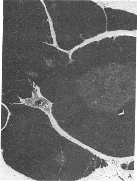
Thymic size and T-cell function decrease with age, and it has not yet been possible to totally reverse this thymic atrophy and completely restore T-cell-dependent immune functions. In this study, GH3 pituitary adenoma cells, which secrete growth hormone and prolactin, were implanted subcutaneously into 16- and 22-month-old female Wistar-Furth rats and the rats were sacrificed approximately 2 months later. Only thymic remnants were detected in aged, non-implanted rats, but thymus glands were found in both the 18- and the 24-month-old rats that had been implanted with GH3 cells. Thymus glands from the GH3-implanted 18-month-old rats contained distinct cortical thymocytes and medullary epithelial cells. Depending on the concentration of phytohemagglutinin or concanavalin A, T-cell proliferative responses of splenocytes from these implanted rats were 2- to 5-fold greater than those of 18-month-old controls. At the optimal concentration of mitogen, proliferative responses to either lectin could be restored to those levels observed in splenocytes from 3-month-old Wistar-Furth females. Thymus glands from 24-month-old GH3-implanted rats contained more cortical thymocytes and fewer fat vacuoles than controls, but they were not totally reconstituted. No significant lectin-induced T-cell proliferative responses or IL-2 secretion were detected in 24-month-old control rats, but splenocytes from GH3-implanted rats showed augmented T-cell proliferative responses and increased synthesis of IL-2. Fluorescence-activated cell-sorter analysis of thymocytes revealed that 24-month-old rats implanted with GH3 cells had a higher proportion of lymphocytes with the Thy-1.1 and helper-T-cell phenotypes. These data show that it is possible to regenerate normal thymic tissue in situ and reverse the natural loss in cell-mediated immunity that occurs with aging.
胸腺大小和T细胞功能随年龄增长而下降,目前尚无法完全逆转这种胸腺萎缩并完全恢复T细胞依赖性免疫功能。在本研究中,将分泌生长激素和催乳素的GH3垂体腺瘤细胞皮下植入16和22月龄的雌性Wistar-Furth大鼠体内,大约2个月后处死大鼠。在未植入的老年大鼠中仅检测到胸腺残余,但在植入GH3细胞的18和24月龄大鼠中均发现了胸腺。植入GH3细胞的18月龄大鼠的胸腺含有明显的皮质胸腺细胞和髓质上皮细胞。根据植物血凝素或刀豆球蛋白A的浓度,这些植入大鼠的脾细胞的T细胞增殖反应比18月龄对照大鼠高2至5倍。在有丝分裂原的最佳浓度下,对任何一种凝集素的增殖反应都可以恢复到3月龄Wistar-Furth雌性大鼠脾细胞中观察到的水平。植入GH3细胞的24月龄大鼠的胸腺比对照含有更多的皮质胸腺细胞和更少的脂肪空泡,但它们并未完全重建。在24月龄对照大鼠中未检测到明显的凝集素诱导的T细胞增殖反应或IL-2分泌,但植入GH3细胞的大鼠的脾细胞显示出增强的T细胞增殖反应和IL-2合成增加。胸腺细胞的荧光激活细胞分选分析显示,植入GH3细胞的24月龄大鼠中具有Thy-1.1和辅助性T细胞表型的淋巴细胞比例更高。这些数据表明,有可能在原位再生正常胸腺组织,并逆转衰老过程中发生的细胞介导免疫的自然丧失。