O'Boyle Nicky, Sutherland Erin, Berry Catherine C, Davies Robert L
Institute of Infection, Immunity and Inflammation, College of Medical, Veterinary and Life Sciences, University of Glasgow, Glasgow, United Kingdom.
Institute of Molecular Cell and Systems Biology, College of Medical, Veterinary and Life Sciences, University of Glasgow, Glasgow, United Kingdom.
PLoS One. 2017 Jul 26;12(7):e0181583. doi: 10.1371/journal.pone.0181583. eCollection 2017.
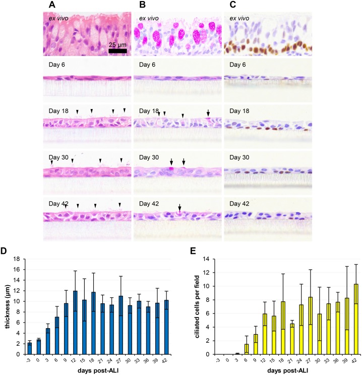
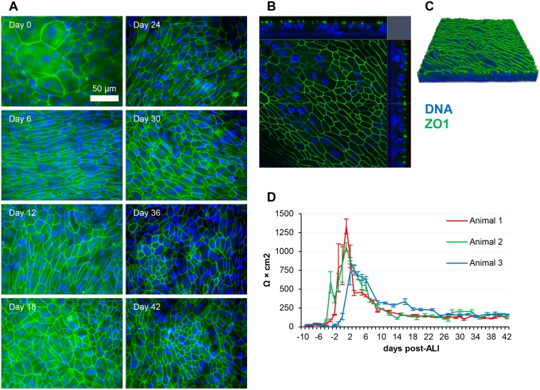
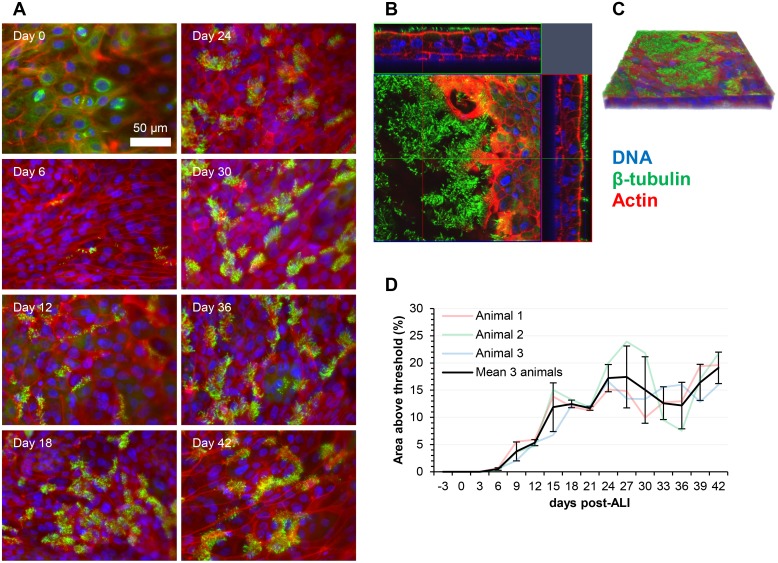
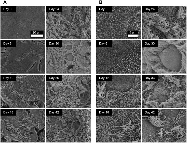
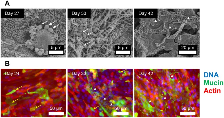
The respiratory tract and lungs are subject to diverse pathologies with wide-ranging implications for both human and animal welfare. The development and detailed characterization of cell culture models for studying such forms of disease is of critical importance. In recent years the use of air-liquid interface (ALI)-cultured airway epithelial cells has increased markedly, as this method of culture results in the formation of a highly representative, organotypic in vitro model system. In this study we have expanded on previous knowledge of differentiated ovine tracheal epithelial cells by analysing the progression of differentiation over an extensive time course at an ALI. We observed a pseudo-stratified epithelium with ciliation and a concurrent increase in cell layer thickness from 9 days post-ALI with ciliation approaching a maximum level at day 24. A similar pattern was observed with respect to mucus production with intensely stained PAS-positive cells appearing at day 12. Ultrastructural analysis by SEM confirmed the presence of both ciliated cells and mucus globules on the epithelial surface within this time-frame. Trans-epithelial electrical resistance (TEER) peaked at 1049 Ω × cm2 as the cell layer became confluent, followed by a subsequent reduction as differentiation proceeded and stabilization at ~200 Ω × cm2. Importantly, little deterioration or de-differentiation was observed over the 45 day time-course indicating that the model is suitable for long-term experiments.
呼吸道和肺部会出现多种病变,对人类和动物健康都有广泛影响。开发用于研究此类疾病形式的细胞培养模型并对其进行详细表征至关重要。近年来,气液界面(ALI)培养的气道上皮细胞的使用显著增加,因为这种培养方法可形成高度代表性的体外器官型模型系统。在本研究中,我们通过分析ALI条件下分化在较长时间过程中的进展,扩展了对分化的绵羊气管上皮细胞的先前认识。我们观察到形成了具有纤毛的假复层上皮,并且从ALI后9天开始细胞层厚度同时增加,纤毛在第24天接近最大水平。在黏液产生方面也观察到类似模式,在第12天出现强染色的PAS阳性细胞。扫描电子显微镜(SEM)的超微结构分析证实,在此时间范围内上皮表面存在纤毛细胞和黏液小球。随着细胞层汇合,跨上皮电阻(TEER)在1049 Ω×cm²达到峰值,随后随着分化进行而降低,并稳定在约200 Ω×cm²。重要的是,在45天的时间过程中几乎未观察到退化或去分化现象,这表明该模型适用于长期实验。