• 文献检索
  • 文档翻译
  • 深度研究
  • 学术资讯
  • Suppr Zotero 插件Zotero 插件
  • 邀请有礼
  • 套餐&价格
  • 历史记录
应用&插件
Suppr Zotero 插件Zotero 插件浏览器插件Mac 客户端Windows 客户端微信小程序
定价
高级版会员购买积分包购买API积分包
服务
文献检索文档翻译深度研究API 文档MCP 服务
关于我们
关于 Suppr公司介绍联系我们用户协议隐私条款
关注我们

Suppr 超能文献

核心技术专利:CN118964589B侵权必究
粤ICP备2023148730 号-1Suppr @ 2026

文献检索

告别复杂PubMed语法,用中文像聊天一样搜索,搜遍4000万医学文献。AI智能推荐,让科研检索更轻松。

立即免费搜索

文件翻译

保留排版,准确专业,支持PDF/Word/PPT等文件格式,支持 12+语言互译。

免费翻译文档

深度研究

AI帮你快速写综述,25分钟生成高质量综述,智能提取关键信息,辅助科研写作。

立即免费体验

爬行动物心脏体静脉干的形态功能特征。

Morpho-functional characterization of the systemic venous pole of the reptile heart.

机构信息

Department of Medical Biology, Academic Medical Center, University of Amsterdam, Amsterdam, The Netherlands.

Department of Bioscience, Zoophysiology, Aarhus University, Aarhus, Denmark.

出版信息

Sci Rep. 2017 Jul 27;7(1):6644. doi: 10.1038/s41598-017-06291-z.

DOI:10.1038/s41598-017-06291-z
PMID:28751678
原文链接:https://pmc.ncbi.nlm.nih.gov/articles/PMC5532247/
Abstract

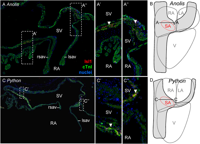
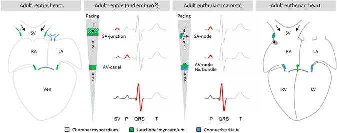
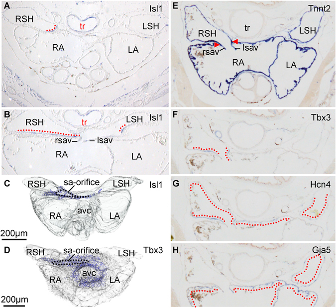
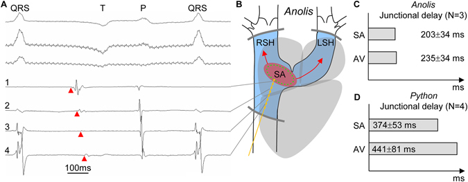
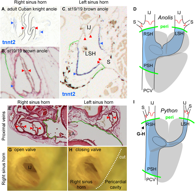
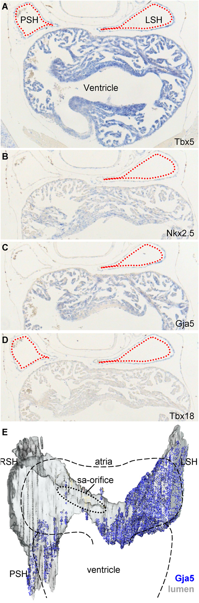
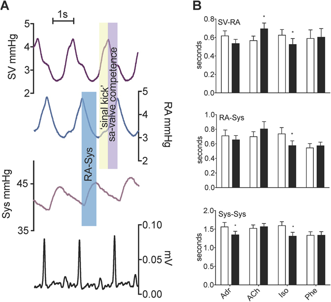
Mammals evolved from reptile-like ancestors, and while the mammalian heart is driven by a distinct sinus node, a sinus node is not apparent in reptiles. We characterized the myocardial systemic venous pole, the sinus venosus, in reptiles to identify the dominant pacemaker and to assess whether the sinus venosus remodels and adopts an atrium-like phenotype as observed in mammals. Anolis lizards had an extensive sinus venosus of myocardium expressing Tbx18. A small sub-population of cells encircling the sinuatrial junction expressed Isl1, Bmp2, Tbx3, and Hcn4, homologues of genes marking the mammalian sinus node. Electrical mapping showed that hearts of Anolis lizards and Python snakes were driven from the sinuatrial junction. The electrical impulse was delayed between the sinus venosus and the right atrium, allowing the sinus venosus to contract and aid right atrial filling. In proximity of the systemic veins, the Anolis sinus venosus expressed markers of the atrial phenotype Nkx2-5 and Gja5. In conclusion, the reptile heart is driven by a pacemaker region with an expression signature similar to that of the immature sinus node of mammals. Unlike mammals, reptiles maintain a sinuatrial delay of the impulse, allowing the partly atrialized sinus venosus to function as a chamber.

摘要

哺乳动物由类似爬行动物的祖先进化而来,虽然哺乳动物的心脏由独特的窦房结驱动,但在爬行动物中并不明显。我们对爬行动物的心肌系统静脉极(窦房静脉)进行了特征描述,以确定主导起搏点,并评估窦房静脉是否会像在哺乳动物中那样发生重塑并采用类似心房的表型。变色龙蜥蜴的窦房静脉有大量表达 Tbx18 的心肌。一小部分细胞环绕着窦房结表达了 Isl1、Bmp2、Tbx3 和 Hcn4,这些基因是标记哺乳动物窦房结的同源基因。电映射显示,变色龙蜥蜴和蟒蛇的心脏由窦房结驱动。电脉冲在窦房静脉和右心房之间延迟,使窦房静脉收缩并有助于右心房充盈。在靠近体静脉的地方,变色龙的窦房静脉表达了心房表型的标志物 Nkx2-5 和 Gja5。总之,爬行动物的心脏由一个具有与哺乳动物未成熟窦房结相似表达特征的起搏区驱动。与哺乳动物不同的是,爬行动物的冲动在窦房结处存在延迟,使部分心房化的窦房静脉能够作为一个腔室发挥作用。

https://cdn.ncbi.nlm.nih.gov/pmc/blobs/7170/5532247/cd7ae205f202/41598_2017_6291_Fig7_HTML.jpg
https://cdn.ncbi.nlm.nih.gov/pmc/blobs/7170/5532247/d542c845ef7e/41598_2017_6291_Fig1_HTML.jpg
https://cdn.ncbi.nlm.nih.gov/pmc/blobs/7170/5532247/86fbb7f554f8/41598_2017_6291_Fig2_HTML.jpg
https://cdn.ncbi.nlm.nih.gov/pmc/blobs/7170/5532247/1d3bfe7bb7b0/41598_2017_6291_Fig3_HTML.jpg
https://cdn.ncbi.nlm.nih.gov/pmc/blobs/7170/5532247/34428744d76a/41598_2017_6291_Fig4_HTML.jpg
https://cdn.ncbi.nlm.nih.gov/pmc/blobs/7170/5532247/b9b164f394a0/41598_2017_6291_Fig5_HTML.jpg
https://cdn.ncbi.nlm.nih.gov/pmc/blobs/7170/5532247/383e7e4d04f8/41598_2017_6291_Fig6_HTML.jpg
https://cdn.ncbi.nlm.nih.gov/pmc/blobs/7170/5532247/cd7ae205f202/41598_2017_6291_Fig7_HTML.jpg

相似文献

1
Morpho-functional characterization of the systemic venous pole of the reptile heart.爬行动物心脏体静脉干的形态功能特征。
Sci Rep. 2017 Jul 27;7(1):6644. doi: 10.1038/s41598-017-06291-z.
2
Molecular pathway for the localized formation of the sinoatrial node.窦房结局部形成的分子途径。
Circ Res. 2007 Feb 16;100(3):354-62. doi: 10.1161/01.RES.0000258019.74591.b3. Epub 2007 Jan 18.
3
The sinus venosus progenitors separate and diversify from the first and second heart fields early in development.窦房静脉祖细胞在发育早期从第一和第二心区分离并多样化。
Cardiovasc Res. 2010 Jul 1;87(1):92-101. doi: 10.1093/cvr/cvq033. Epub 2010 Jan 28.
4
The sinus venosus myocardium contributes to the atrioventricular canal: potential role during atrioventricular node development?静脉窦心肌对房室管有贡献:在房室结发育过程中的潜在作用?
J Cell Mol Med. 2015 Jun;19(6):1375-89. doi: 10.1111/jcmm.12525. Epub 2015 Mar 6.
5
Identification and functional characterization of cardiac pacemaker cells in zebrafish.鉴定和功能表征斑马鱼的心脏起搏细胞。
PLoS One. 2012;7(10):e47644. doi: 10.1371/journal.pone.0047644. Epub 2012 Oct 16.
6
Structure and function of the hearts of lizards and snakes.蜥蜴和蛇类心脏的结构与功能。
Biol Rev Camb Philos Soc. 2014 May;89(2):302-36. doi: 10.1111/brv.12056. Epub 2013 Sep 2.
7
Morphological and functional aspects of reptilian ultimobranchial gland.爬行动物后鳃腺的形态学和功能方面
Anat Histol Embryol. 1998 Dec;27(6):359-64. doi: 10.1111/j.1439-0264.1998.tb00208.x.
8
Molecular analysis of patterning of conduction tissues in the developing human heart.人类心脏发育中传导组织模式的分子分析。
Circ Arrhythm Electrophysiol. 2011 Aug;4(4):532-42. doi: 10.1161/CIRCEP.111.963421. Epub 2011 May 16.
9
A common Shox2-Nkx2-5 antagonistic mechanism primes the pacemaker cell fate in the pulmonary vein myocardium and sinoatrial node.一种常见的Shox2-Nkx2-5拮抗机制决定了肺静脉心肌和窦房结中起搏细胞的命运。
Development. 2015 Jul 15;142(14):2521-32. doi: 10.1242/dev.120220. Epub 2015 Jul 2.
10
Sinus venosus incorporation: contentious issues and operational criteria for developmental and evolutionary studies.静脉窦融合:发育和进化研究中的争议问题和操作标准。
J Anat. 2019 May;234(5):583-591. doi: 10.1111/joa.12962. Epub 2019 Mar 12.

引用本文的文献

1
Electrophysiological evidence for the right atrioventricular pacemaker ring in the reptile heart.爬行动物心脏右房室起搏环的电生理证据。
J Comp Physiol B. 2025 Apr;195(2):181-190. doi: 10.1007/s00360-025-01612-5. Epub 2025 Mar 28.
2
The simple morphology of the sunfish heart.太阳鱼心脏的简单形态。
J Anat. 2025 Jun;246(6):1034-1041. doi: 10.1111/joa.14198. Epub 2024 Dec 20.
3
Anatomy of the heart of the leatherback turtle.棱皮龟心脏解剖结构。

本文引用的文献

1
Misexpression of Tbx18 in cardiac chambers of fetal mice interferes with chamber-specific developmental programs but does not induce a pacemaker-like gene signature.Tbx18在胎鼠心腔中的错误表达会干扰心腔特异性发育程序,但不会诱导起搏器样基因特征。
J Mol Cell Cardiol. 2016 Aug;97:140-9. doi: 10.1016/j.yjmcc.2016.05.004. Epub 2016 May 11.
2
RNA sequencing of mouse sinoatrial node reveals an upstream regulatory role for Islet-1 in cardiac pacemaker cells.小鼠窦房结的RNA测序揭示了Islet-1在心脏起搏细胞中的上游调控作用。
Circ Res. 2015 Feb 27;116(5):797-803. doi: 10.1161/CIRCRESAHA.116.305913. Epub 2015 Jan 26.
3
J Anat. 2022 Aug;241(2):535-544. doi: 10.1111/joa.13670. Epub 2022 Apr 12.
4
Why are you talking with snakes? To get new evolutionary insights in cardiac electrophysiology!你为什么要和蛇说话?是为了在心脏电生理学方面获得新的进化见解!
J Gen Physiol. 2022 Apr 4;154(4). doi: 10.1085/jgp.202113060. Epub 2022 Mar 17.
5
Anatomy of the heart with the highest heart rate.心脏解剖图与最大心率。
J Anat. 2022 Jul;241(1):173-190. doi: 10.1111/joa.13640. Epub 2022 Feb 6.
6
A characterization of the electrophysiological properties of the cardiomyocytes from ventricle, atrium and sinus venosus of the snake heart.蛇心脏心室、心房和静脉窦心肌细胞电生理特性的表征
J Comp Physiol B. 2020 Jan;190(1):63-73. doi: 10.1007/s00360-019-01253-5. Epub 2019 Dec 18.
7
Reptiles as a Model System to Study Heart Development.爬行动物作为研究心脏发育的模型系统。
Cold Spring Harb Perspect Biol. 2020 May 1;12(5):a037226. doi: 10.1101/cshperspect.a037226.
8
Low incidence of atrial septal defects in nonmammalian vertebrates.非哺乳动物脊椎动物中心房间隔缺损的发生率较低。
Evol Dev. 2020 May;22(3):241-256. doi: 10.1111/ede.12322. Epub 2019 Oct 9.
9
Smooth Muscle in Cardiac Chambers is Common in Turtles and Extensive in the Emydid Turtle, Trachemys scripta.龟类心室中的平滑肌很常见,而在拟鳄龟(Trachemys scripta)中则非常广泛。
Anat Rec (Hoboken). 2020 May;303(5):1327-1336. doi: 10.1002/ar.24257. Epub 2019 Oct 10.
10
Sinus venosus incorporation: contentious issues and operational criteria for developmental and evolutionary studies.静脉窦融合:发育和进化研究中的争议问题和操作标准。
J Anat. 2019 May;234(5):583-591. doi: 10.1111/joa.12962. Epub 2019 Mar 12.
Physiological contractility of cardiomyocytes in the wall of mouse and rat azygos vein.
心肌细胞在鼠和大鼠奇静脉壁中的生理收缩性。
Am J Physiol Cell Physiol. 2014 Apr 1;306(7):C697-704. doi: 10.1152/ajpcell.00004.2014. Epub 2014 Jan 29.
4
Structure and function of the hearts of lizards and snakes.蜥蜴和蛇类心脏的结构与功能。
Biol Rev Camb Philos Soc. 2014 May;89(2):302-36. doi: 10.1111/brv.12056. Epub 2013 Sep 2.
5
Phylogeny and effects of anoxia on hyperpolarization-activated cyclic nucleotide-gated channel gene expression in the heart of a primitive chordate, the Pacific hagfish (Eptatretus stoutii).在原始脊索动物——太平洋盲鳗(Eptatretus stoutii)的心脏中,缺氧对超极化激活环核苷酸门控通道基因表达的进化和影响。
J Exp Biol. 2013 Dec 1;216(Pt 23):4462-72. doi: 10.1242/jeb.094912. Epub 2013 Aug 30.
6
Development of the hearts of lizards and snakes and perspectives to cardiac evolution.蜥蜴和蛇类心脏的发育与心脏演化的展望。
PLoS One. 2013 Jun 5;8(6):e63651. doi: 10.1371/journal.pone.0063651. Print 2013.
7
Structure, function and clinical relevance of the cardiac conduction system, including the atrioventricular ring and outflow tract tissues.心脏传导系统的结构、功能和临床相关性,包括房室环和流出道组织。
Pharmacol Ther. 2013 Aug;139(2):260-88. doi: 10.1016/j.pharmthera.2013.04.010. Epub 2013 Apr 20.
8
Early mesodermal cues assign avian cardiac pacemaker fate potential in a tertiary heart field.早期中胚层线索在三级心脏场中指定禽类心脏起搏命运潜能。
Science. 2013 May 10;340(6133):744-8. doi: 10.1126/science.1232877. Epub 2013 Mar 21.
9
Islet1 is a direct transcriptional target of the homeodomain transcription factor Shox2 and rescues the Shox2-mediated bradycardia.胰岛 1 是同源域转录因子 Shox2 的直接转录靶标,并挽救了 Shox2 介导的心动过缓。
Basic Res Cardiol. 2013 Mar;108(2):339. doi: 10.1007/s00395-013-0339-z. Epub 2013 Mar 1.
10
Discontinuous thoracic venous cardiomyocytes and heart exhibit synchronized developmental switch of troponin isoforms.间断性胸静脉心肌细胞和心脏表现出肌钙蛋白同工型的发育同步转换。
FEBS J. 2013 Feb;280(3):880-91. doi: 10.1111/febs.12076. Epub 2013 Jan 2.