Radziuviene Gedmante, Rasmusson Allan, Augulis Renaldas, Lesciute-Krilaviciene Daiva, Laurinaviciene Aida, Clim Eduard, Laurinavicius Arvydas
National Center of Pathology, Affiliate of Vilnius University Hospital Santariskiu Clinics, P. Baublio 5, LT-08406 Vilnius, Lithuania.
Faculty of Natural Sciences, Vilnius University, M. K. Ciurlionio 27, LT-03103 Vilnius, Lithuania.
Biomed Res Int. 2017;2017:2321916. doi: 10.1155/2017/2321916. Epub 2017 May 28.
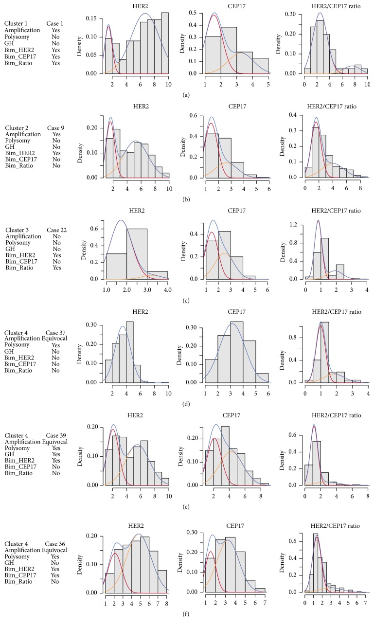
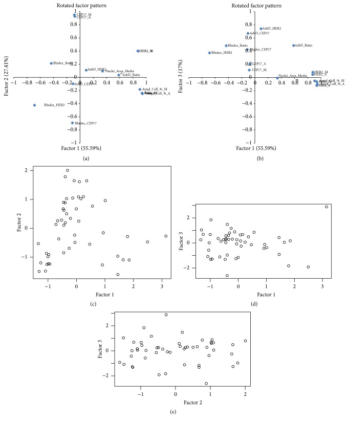
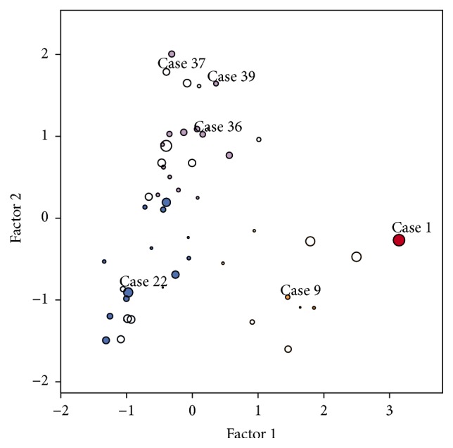
Human epidermal growth factor receptor 2 gene- (HER2-) targeted therapy for breast cancer relies primarily on HER2 overexpression established by immunohistochemistry (IHC) with borderline cases being further tested for amplification by fluorescence in situ hybridization (FISH). Manual interpretation of HER2 FISH is based on a limited number of cells and rather complex definitions of equivocal, polysomic, and genetically heterogeneous (GH) cases. Image analysis (IA) can extract high-capacity data and potentially improve HER2 testing in borderline cases. We investigated statistically derived indicators of HER2 heterogeneity in HER2 FISH data obtained by automated IA of 50 IHC borderline (2+) cases of invasive ductal breast carcinoma. Overall, IA significantly underestimated the conventional HER2, CEP17 counts, and HER2/CEP17 ratio; however, it collected more amplified cells in some cases below the lower limit of GH definition by manual procedure. Indicators for amplification, polysomy, and bimodality were extracted by factor analysis and allowed clustering of the tumors into amplified, nonamplified, and equivocal/polysomy categories. The bimodality indicator provided independent cell diversity characteristics for all clusters. Tumors classified as bimodal only partially coincided with the conventional GH heterogeneity category. We conclude that automated high-capacity nonselective tumor cell assay can generate evidence-based HER2 intratumor heterogeneity indicators to refine GH definitions.
人表皮生长因子受体2基因(HER2)靶向治疗乳腺癌主要依赖免疫组织化学(IHC)确定的HER2过表达,临界病例需进一步通过荧光原位杂交(FISH)检测扩增情况。HER2 FISH的人工判读基于有限数量的细胞以及对意义不明确、多体性和基因异质性(GH)病例相当复杂的定义。图像分析(IA)可以提取大容量数据,并有可能改善临界病例的HER2检测。我们对50例浸润性导管癌IHC临界(2+)病例通过自动IA获得的HER2 FISH数据中HER2异质性的统计学衍生指标进行了研究。总体而言,IA显著低估了传统的HER2、CEP17计数和HER2/CEP17比值;然而,在一些按人工流程低于GH定义下限的病例中,它收集到了更多扩增细胞。通过因子分析提取了扩增、多体性和双峰性的指标,并可将肿瘤聚类为扩增、未扩增以及意义不明确/多体性类别。双峰性指标为所有聚类提供了独立的细胞多样性特征。分类为双峰的肿瘤仅部分与传统的GH异质性类别相符。我们得出结论,自动化大容量非选择性肿瘤细胞检测可以生成基于证据的HER2肿瘤内异质性指标,以完善GH定义。