Unité de Recherche en Santé des Populations, Centre de Recherche du CHU de Québec, Hôpital du St-Sacrement, 1050 Chemin Ste-Foy, Quebec City, QC G1S4L8, Canada.
Diagn Pathol. 2013 Feb 4;8:17. doi: 10.1186/1746-1596-8-17.
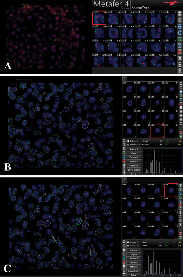
Amplification of the human epidermal growth factor receptor 2 (HER2) is a prognostic marker for poor clinical outcome and a predictive marker for therapeutic response to targeted therapies in breast cancer patients. With the introduction of anti-HER2 therapies, accurate assessment of HER2 status has become essential. Fluorescence in situ hybridization (FISH) is a widely used technique for the determination of HER2 status in breast cancer. However, the manual signal enumeration is time-consuming. Therefore, several companies like MetaSystem have developed automated image analysis software. Some of these signal enumeration software employ the so called "tile-sampling classifier", a programming algorithm through which the software quantifies fluorescent signals in images on the basis of square tiles of fixed dimensions. Considering that the size of tile does not always correspond to the size of a single tumor cell nucleus, some users argue that this analysis method might not completely reflect the biology of cells. For that reason, MetaSystems has developed a new classifier which is able to recognize nuclei within tissue sections in order to determine the HER2 amplification status on nuclei basis. We call this new programming algorithm "nuclei-sampling classifier". In this study, we evaluated the accuracy of the "nuclei-sampling classifier" in determining HER2 gene amplification by FISH in nuclei of breast cancer cells. To this aim, we randomly selected from our cohort 64 breast cancer specimens (32 nonamplified and 32 amplified) and we compared results obtained through manual scoring and through this new classifier. The new classifier automatically recognized individual nuclei. The automated analysis was followed by an optional human correction, during which the user interacted with the software in order to improve the selection of cell nuclei automatically selected. Overall concordance between manual scoring and automated nuclei-sampling analysis was 98.4% (100% for nonamplified cases and 96.9% for amplified cases). However, after human correction, concordance between the two methods was 100%. We conclude that the nuclei-based classifier is a new available tool for automated quantitative HER2 FISH signals analysis in nuclei in breast cancer specimen and it can be used for clinical purposes.
人类表皮生长因子受体 2(HER2)的扩增是乳腺癌患者临床预后不良的标志物,也是靶向治疗疗效的预测标志物。随着抗 HER2 治疗的引入,准确评估 HER2 状态变得至关重要。荧光原位杂交(FISH)是一种广泛用于确定乳腺癌 HER2 状态的技术。然而,手动信号计数既耗时又繁琐。因此,像 MetaSystem 这样的几家公司已经开发了自动化图像分析软件。这些信号计数软件中的一些采用了所谓的“平铺采样分类器”,这是一种编程算法,通过该算法,软件可以根据固定尺寸的正方形平铺来量化图像中的荧光信号。考虑到平铺的大小并不总是与单个肿瘤细胞核的大小相对应,一些用户认为这种分析方法可能无法完全反映细胞的生物学特性。出于这个原因,MetaSystems 开发了一种新的分类器,能够识别组织切片中的细胞核,以便根据细胞核基础确定 HER2 扩增状态。我们称这个新的编程算法为“细胞核采样分类器”。在这项研究中,我们评估了“细胞核采样分类器”在通过 FISH 确定乳腺癌细胞核中 HER2 基因扩增方面的准确性。为此,我们从我们的队列中随机选择了 64 个乳腺癌标本(32 个未扩增和 32 个扩增),并将通过手动评分和这个新分类器获得的结果进行了比较。新分类器自动识别单个细胞核。自动化分析后,可以进行可选的人工校正,在此期间,用户与软件进行交互,以改进自动选择的细胞核的选择。手动评分和自动细胞核采样分析之间的总体一致性为 98.4%(非扩增病例为 100%,扩增病例为 96.9%)。然而,经过人工校正后,两种方法之间的一致性达到了 100%。我们得出结论,基于细胞核的分类器是一种新的可用于自动定量分析乳腺癌标本细胞核中 HER2 FISH 信号的工具,可用于临床目的。