Kuriyama Takumi, Sakai Nobuyuki, Beppu Mikiya, Sakai Chiaki, Imamura Hirotoshi, Kojima Iwao, Masago Katsuhiro, Katakami Nobuyuki
1 Division of Radiological Technology, Institute of Biomedical Research and Innovation, Kobe, Japan.
2 Division of Neuro-endovascular Therapy, Institute of Biomedical Research and Innovation, Kobe, Japan.
J Int Med Res. 2018 Jan;46(1):464-474. doi: 10.1177/0300060517715165. Epub 2017 Jul 31.
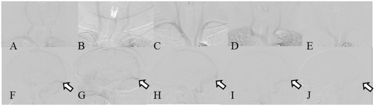
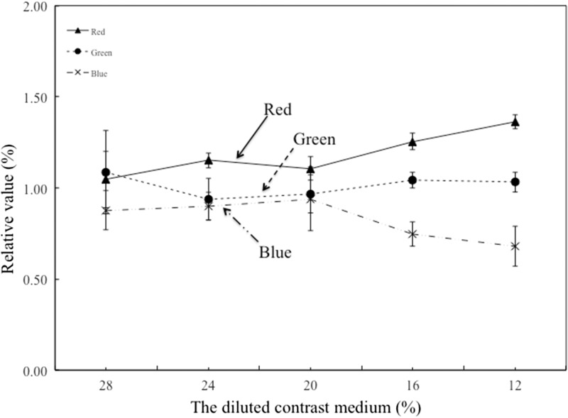
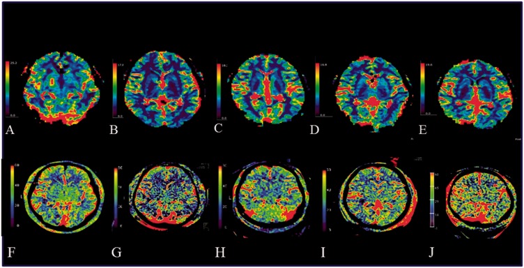
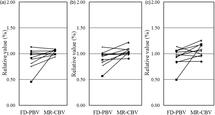
Objective Similar to perfusion studies after acute ischemic stroke, measuring cerebral blood volume (CBV) via C-arm computed tomography before and after therapeutic interventions may help gauge subsequent revascularization. We tested serial dilutions of intra-arterial injectable contrast medium (CM) to determine the optimal CM concentration for quantifying parenchymal blood volume by flat-panel detector imaging (FD-PBV). Methods CM was diluted via saline power injector, instituting time delays for FD-PBV studies. A red/green/blue (RGB) color scale was employed to quantify/compare FD-PBV and magnetic resonance-derived CBV (MRCBV). Results Contrast values of right and left common carotid arteries did not differ significantly at CM dilutions of ≥20%. RGB analysis of FD-PBV imaging (relative to MR-CVB), showed CM dilution altered the colors (by 16%), increasing red and decreasing blue ratios. Conclusion Diluting CM to 20% resulted in no laterality differential of FD-PBV imaging, with left/right quantitative ratios approaching 1.1 (optimal for clinical use).
目的 与急性缺血性卒中后的灌注研究类似,在治疗干预前后通过C形臂计算机断层扫描测量脑血容量(CBV)可能有助于评估随后的血管再通情况。我们测试了动脉内注射造影剂(CM)的系列稀释液,以确定通过平板探测器成像(FD-PBV)定量实质血容量的最佳CM浓度。方法 通过盐水动力注射器稀释CM,为FD-PBV研究设置时间延迟。采用红/绿/蓝(RGB)色标来量化/比较FD-PBV和磁共振衍生的CBV(MRCBV)。结果 在CM稀释度≥20%时,左右颈总动脉的对比值无显著差异。FD-PBV成像的RGB分析(相对于MR-CVB)显示,CM稀释改变了颜色(改变了16%),红色比例增加,蓝色比例降低。结论 将CM稀释至20%可使FD-PBV成像无左右差异差异,左右定量比率接近1.1(临床使用的最佳值)。