Yoo Hong Yeol, Choi Jaewoo, Kim Jongjin, Chai Young Jun, Shin Rumi, Ahn Hye Seong, Lim Chang-Sup, Lee Hae Won, Hwang Ki-Tae, Jung In Mok, Chung Jung Kee, Heo Seung Chul
Department of Surgery, Seoul Metropolitan Government - Seoul National University Boramae Medical Center, Seoul, Korea.
Ann Coloproctol. 2017 Jun;33(3):99-105. doi: 10.3393/ac.2017.33.3.99. Epub 2017 Jun 30.
The preoperative diagnosis of acute appendicitis is often challenging. Sometimes, pathologic results of the appendix embarrass or confuse surgeons. Therefore, more and more imaging studies are being performed to increase the accuracy of appendicitis diagnoses preoperatively. However, data on the effect of this increase in preoperative imaging studies on diagnostic accuracy are limited. We performed this study to explore unexpected appendiceal pathologies and to delineate the role of preoperative imaging studies in the diagnosis of acute appendicitis.
The medical records of 4,673 patients who underwent an appendectomy for assumed appendicitis between 1997 and 2012 were reviewed retrospectively. Pathological results and preoperative imaging studies were surveyed, and the frequencies of pathological results and preoperative imaging studies were investigated.
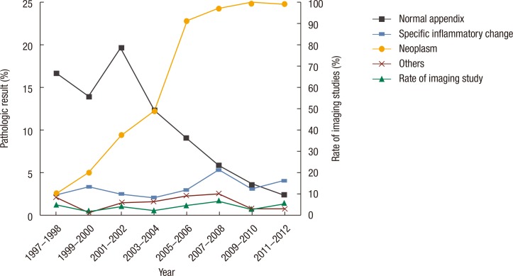
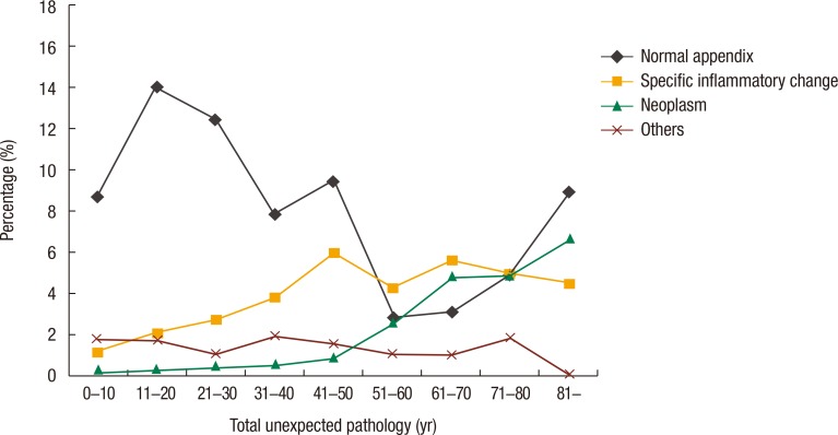
The overall rate of pathology compatible with acute appendicitis was 84.4%. Unexpected pathological findings, such as normal histology, specific inflammations other than acute appendicitis, neoplastic lesions, and other pathologies, comprised 9.6%, 3.3%, 1.2%, and 1.5%, respectively. The rate of unexpected pathological results was significantly reduced because of the increase in preoperative imaging studies. The decrease in normal appendices contributed the most to the reduction while other unexpected pathologies did not change significantly despite the increased use of imaging studies. This decrease in normal appendices was significant in both male and female patients under the age of 60 years, but the differences in females were more prominent.
Unexpected appendiceal pathologies comprised 15.6% of the cases. Preoperative imaging studies reduced them by decreasing the negative appendectomy rate of patients with normal appendices.
急性阑尾炎的术前诊断常常具有挑战性。有时,阑尾的病理结果会让外科医生感到尴尬或困惑。因此,越来越多的影像学检查被用于提高术前阑尾炎诊断的准确性。然而,关于术前影像学检查增加对诊断准确性影响的数据有限。我们开展这项研究以探索意外的阑尾病变,并阐明术前影像学检查在急性阑尾炎诊断中的作用。
回顾性分析1997年至2012年间因疑似阑尾炎接受阑尾切除术的4673例患者的病历。调查病理结果和术前影像学检查情况,并研究病理结果和术前影像学检查的频率。
与急性阑尾炎相符的病理总体发生率为84.4%。意外病理发现,如组织学正常、非急性阑尾炎的特异性炎症、肿瘤性病变和其他病变,分别占9.6%、3.3%、1.2%和1.5%。由于术前影像学检查的增加,意外病理结果的发生率显著降低。正常阑尾病例数的减少对这种降低贡献最大,而尽管影像学检查使用增加,其他意外病变并未显著改变。60岁以下男性和女性患者中正常阑尾病例数的减少均显著,但女性中的差异更为突出。
意外的阑尾病变占病例的15.6%。术前影像学检查通过降低正常阑尾患者的阴性阑尾切除率减少了这些病变。