Karolinska Institutet, Department NVS, Center for Alzheimer Research, Division of Neurogeriatrics, Huddinge, Sweden.
Present address: Dainippon Sumitomo Pharma Co., Ltd, Drug Development Research Laboratories, Osaka, Japan.
Alzheimers Res Ther. 2017 Aug 1;9(1):57. doi: 10.1186/s13195-017-0279-1.
Increased levels of the pathogenic amyloid β-peptide (Aβ), released from its precursor by the transmembrane protease γ-secretase, are found in Alzheimer disease (AD) brains. Interestingly, monoamine oxidase B (MAO-B) activity is also increased in AD brain, but its role in AD pathogenesis is not known. Recent neuroimaging studies have shown that the increased MAO-B expression in AD brain starts several years before the onset of the disease. Here, we show a potential connection between MAO-B, γ-secretase and Aβ in neurons.
MAO-B immunohistochemistry was performed on postmortem human brain. Affinity purification of γ-secretase followed by mass spectrometry was used for unbiased identification of γ-secretase-associated proteins. The association of MAO-B with γ-secretase was studied by coimmunoprecipitation from brain homogenate, and by in-situ proximity ligation assay (PLA) in neurons as well as mouse and human brain sections. The effect of MAO-B on Aβ production and Notch processing in cell cultures was analyzed by siRNA silencing or overexpression experiments followed by ELISA, western blot or FRET analysis. Methodology for measuring relative intraneuronal MAO-B and Aβ42 levels in single cells was developed by combining immunocytochemistry and confocal microscopy with quantitative image analysis.
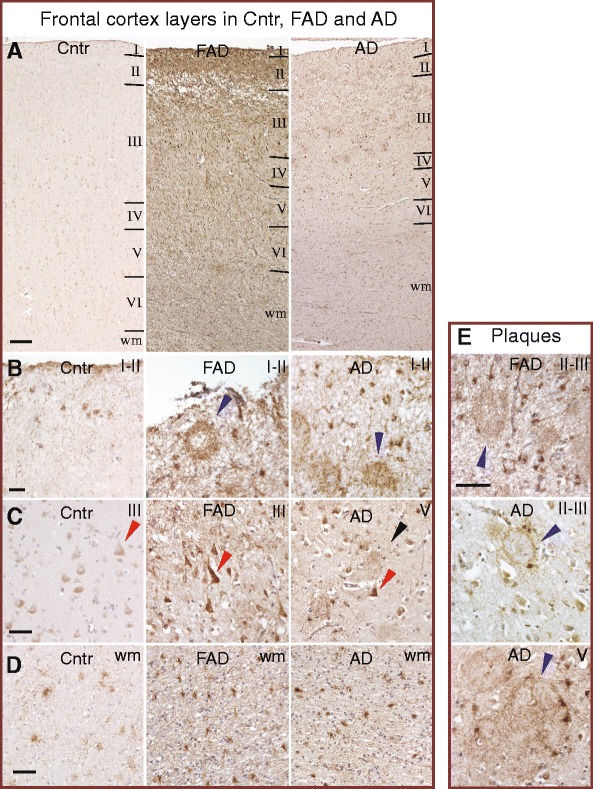
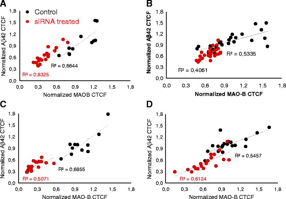
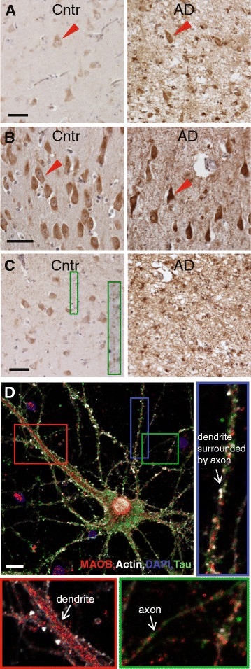
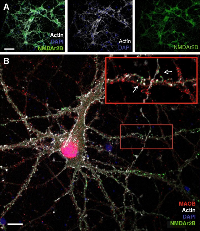
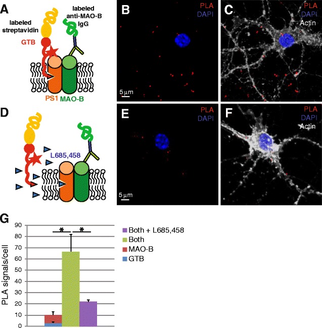
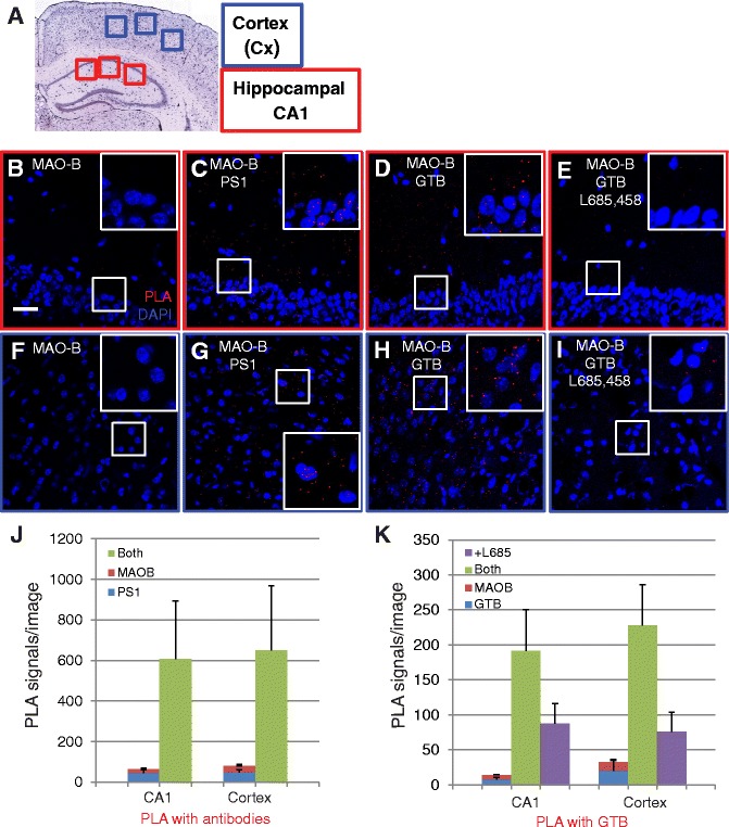
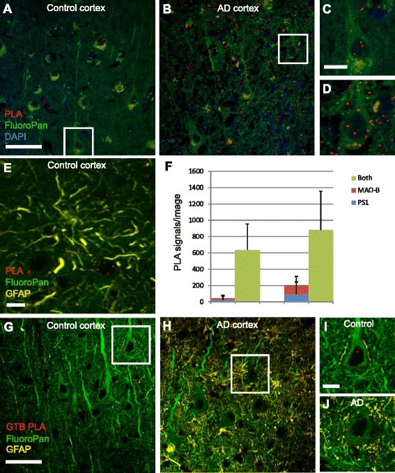
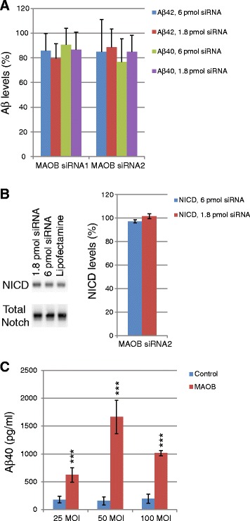
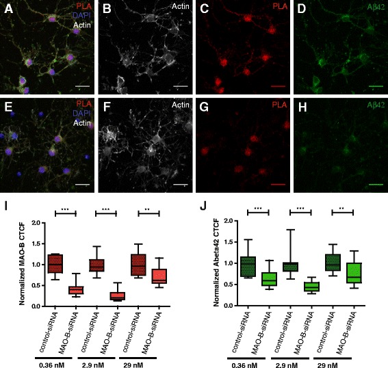
Immunohistochemistry revealed MAO-B staining in neurons in the frontal cortex, hippocampus CA1 and entorhinal cortex in postmortem human brain. Interestingly, the neuronal staining intensity was higher in AD brain than in control brain in these regions. Mass spectrometric data from affinity purified γ-secretase suggested that MAO-B is a γ-secretase-associated protein, which was confirmed by immunoprecipitation and PLA, and a neuronal location of the interaction was shown. Strikingly, intraneuronal Aβ42 levels correlated with MAO-B levels, and siRNA silencing of MAO-B resulted in significantly reduced levels of intraneuronal Aβ42. Furthermore, overexpression of MAO-B enhanced Aβ production.
This study shows that MAO-B levels are increased not only in astrocytes but also in pyramidal neurons in AD brain. The study also suggests that MAO-B regulates Aβ production in neurons via γ-secretase and thereby provides a key to understanding the relationship between MAO-B and AD pathogenesis. Potentially, the γ-secretase/MAO-B association may be a target for reducing Aβ levels using protein-protein interaction breakers.
在阿尔茨海默病(AD)大脑中,淀粉样β肽(Aβ)前体通过跨膜蛋白酶 γ-分泌酶释放,其水平升高。有趣的是,单胺氧化酶 B(MAO-B)的活性在 AD 大脑中也升高,但它在 AD 发病机制中的作用尚不清楚。最近的神经影像学研究表明,AD 大脑中 MAO-B 的表达增加始于疾病发作前数年。在这里,我们展示了神经元中 MAO-B、γ-分泌酶和 Aβ 之间的潜在联系。
对死后人脑进行 MAO-B 免疫组织化学染色。通过亲和纯化 γ-分泌酶,然后进行质谱分析,用于无偏鉴定 γ-分泌酶相关蛋白。通过脑匀浆中的免疫沉淀以及神经元以及小鼠和人脑切片中的原位邻近连接测定(PLA)研究 MAO-B 与 γ-分泌酶的关联。通过 siRNA 沉默或过表达实验,然后通过 ELISA、western blot 或 FRET 分析分析 MAO-B 对 Aβ 产生和 Notch 加工的影响。通过将免疫细胞化学和共聚焦显微镜与定量图像分析相结合,开发了一种测量单个细胞中相对神经元内 MAO-B 和 Aβ42 水平的方法。
免疫组织化学显示 MAO-B 染色存在于死后人脑额皮质、海马 CA1 和内嗅皮质的神经元中。有趣的是,与这些区域的对照大脑相比,AD 大脑中的神经元染色强度更高。亲和纯化 γ-分泌酶的质谱数据表明 MAO-B 是 γ-分泌酶相关蛋白,这通过免疫沉淀和 PLA 得到证实,并显示了相互作用的神经元位置。引人注目的是,神经元内 Aβ42 水平与 MAO-B 水平相关,MAO-B 的 siRNA 沉默导致神经元内 Aβ42 水平显著降低。此外,MAO-B 的过表达增强了 Aβ 的产生。
这项研究表明,MAO-B 水平不仅在星形胶质细胞中升高,而且在 AD 大脑中的锥体细胞中也升高。该研究还表明,MAO-B 通过 γ-分泌酶调节神经元中的 Aβ 产生,从而为理解 MAO-B 与 AD 发病机制之间的关系提供了关键。潜在地,γ-分泌酶/MAO-B 关联可能是使用蛋白质-蛋白质相互作用破坏剂降低 Aβ 水平的靶点。