Granata Vincenza, Fusco Roberta, Setola Sergio Venanzio, Piccirillo Mauro, Leongito Maddalena, Palaia Raffaele, Granata Francesco, Lastoria Secondo, Izzo Francesco, Petrillo Antonella
Vincenza Granata, Roberta Fusco, Sergio Venanzio Setola, Secondo Lastoria, Antonella Petrillo, Department of Diagnostic Imaging, Radiant and Metabolic Therapy, "Istituto Nazionale Tumori IRCCS Fondazione Pascale - IRCCS di Napoli", 80131 Naples, Italy.
World J Gastroenterol. 2017 Jul 14;23(26):4767-4778. doi: 10.3748/wjg.v23.i26.4767.
To report early imaging assessment of ablated area post electrochemotherapy (ECT) in patients with locally advanced pancreatic cancer (LAPC).
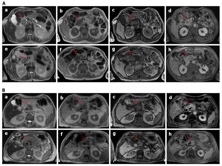
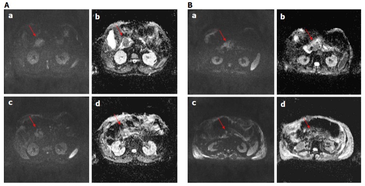
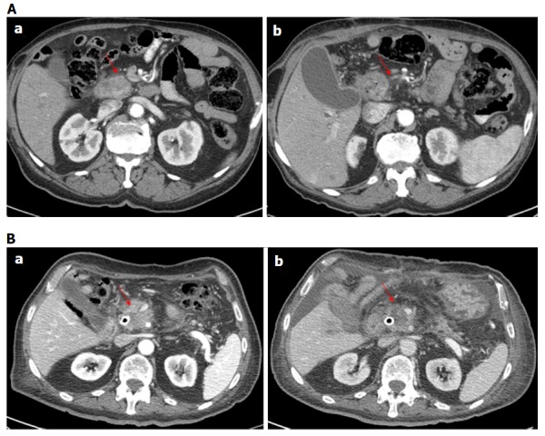
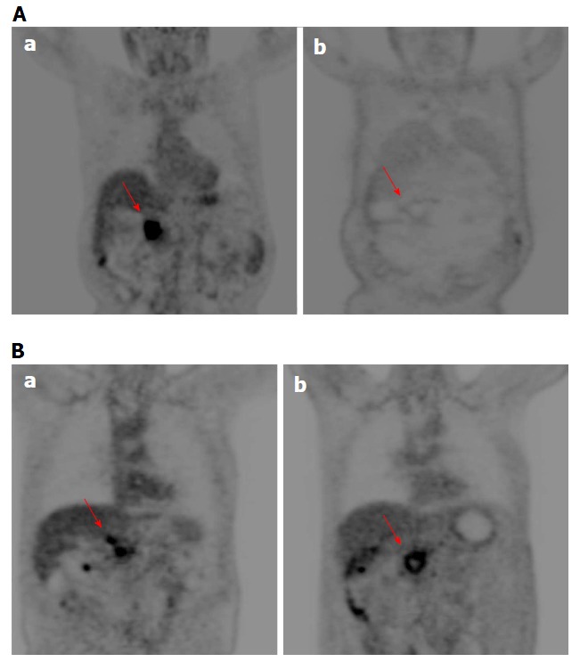
ECT was performed in 19 LAPC patients enrolled in an approved ongoing clinical phase I/II study. Before and after ECT, 18 patients underwent computed tomography (CT) scan, 11 patients underwent morphological and functional magnetic resonance (MR) scan (dynamic contrast enhanced-MRI) calculating wash-in slope (WIS) and wash-out slope (WOS); diffusion weighted imaging calculating pseudo-diffusivity (Dp), perfusion fraction (fp) and tissue diffusivity (Dt); 10 patients underwent positron emission tomography (PET). Response evaluation criteria in solid tumour (RECIST) on MR and CT were used to assess tumour therapy response. Choi on CT, PET response criteria in solid tumors (PERCIST) on PET and functional parameters on MR were used to evaluate treatment response.
For each patient no significant reduction was measurable by CT and MR using RECIST. According Choi criteria a partial response was obtained in 18/18 (100.0%) patients. According PERCIST criteria 6/10 (60.0%) patients showed a partial response, 3/10 (30.0%) stable disease and 1/10 (10.0%) progression disease. Moreover, using functional MR parameters, a significant reduction of viable tumour after ECT can be observed. According ΔWIS and ΔWOS 9/11 (81.8%) patients exhibited a partial response and 2/11 (18.2%) stable disease; 8/11 (72.7%) patients were considered in partial response by ΔDp evaluation and 3/11 (27.3%) in stable disease; according ΔDt 7/11 (63.6%) patients showed a partial response, 1/11 (9.1%) showed progression of disease and 3/11 (27.3%) were stable. Perfusion fraction fp showed a significant reduction after ECT only in four patients. No significant difference was observed after ECT in signal intensity of T1-weighted images and T2-weighted images, and in equilibrium-phase of contrast study, according to χ test was observed. A good correlation was reported between ΔHounsfield unit and Δmaximum standardized uptake value and between Δfp and ΔWOS, with a significant statistically difference ( < 0.05) using Spearman correlation coefficient.
Perfusion and diffusion MR derived parameters, Choi, PERCIST criteria are more performant than morphological MR and CT criteria to assess ECT treatment response.
报告局部晚期胰腺癌(LAPC)患者接受电化学疗法(ECT)后消融区域的早期影像学评估。
对19例参与一项正在进行的获批I/II期临床研究的LAPC患者实施ECT。ECT前后,18例患者接受了计算机断层扫描(CT),11例患者接受了形态学和功能磁共振(MR)扫描(动态对比增强磁共振成像)以计算注入斜率(WIS)和流出斜率(WOS);弥散加权成像以计算伪扩散系数(Dp)、灌注分数(fp)和组织扩散率(Dt);10例患者接受了正电子发射断层扫描(PET)。采用基于MR和CT的实体瘤疗效评价标准(RECIST)评估肿瘤治疗反应。采用基于CT的Choi标准、基于PET的实体瘤PET反应标准(PERCIST)以及基于MR的功能参数评估治疗反应。
根据RECIST标准,通过CT和MR测量,每位患者均未观察到显著缩小。根据Choi标准,18/18(100.0%)例患者获得部分缓解。根据PERCIST标准,6/10(60.0%)例患者显示部分缓解,3/10(30.0%)例疾病稳定,1/10(10.0%)例疾病进展。此外,使用功能MR参数,可观察到ECT后存活肿瘤显著减少。根据ΔWIS和ΔWOS,9/11(81.8%)例患者表现为部分缓解,2/11(18.2%)例疾病稳定;根据ΔDp评估,8/11(72.7%)例患者被认为部分缓解,3/11(27.3%)例疾病稳定;根据ΔDt,7/11(63.6%)例患者显示部分缓解,1/11(9.1%)例显示疾病进展,3/11(27.3%)例疾病稳定。仅4例患者ECT后灌注分数fp显著降低。根据χ检验,ECT后T1加权图像和T2加权图像的信号强度以及对比研究的平衡期未观察到显著差异。据报道,ΔHounsfield单位与Δ最大标准化摄取值之间以及Δfp与ΔWOS之间具有良好的相关性,使用Spearman相关系数具有显著统计学差异(<0.05)。
与形态学MR和CT标准相比,基于灌注和弥散的MR衍生参数、Choi标准、PERCIST标准在评估ECT治疗反应方面表现更优。