Adachi Kyoichi, Mishiro Tomoko, Tanaka Shino, Kinoshita Yoshikazu
Health Center, Shimane Environment and Health Public Corporation, Japan.
The Second Department of Internal Medicine, Shimane University Faculty of Medicine, Japan.
Intern Med. 2017;56(15):1937-1942. doi: 10.2169/internalmedicine.56.8260. Epub 2017 Aug 1.
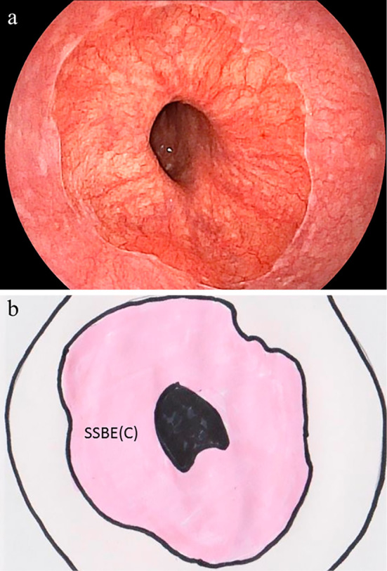
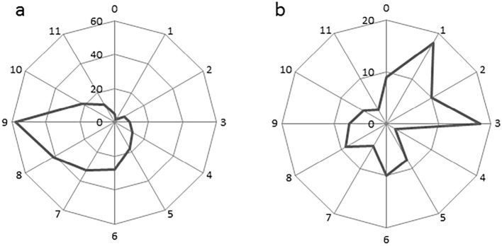
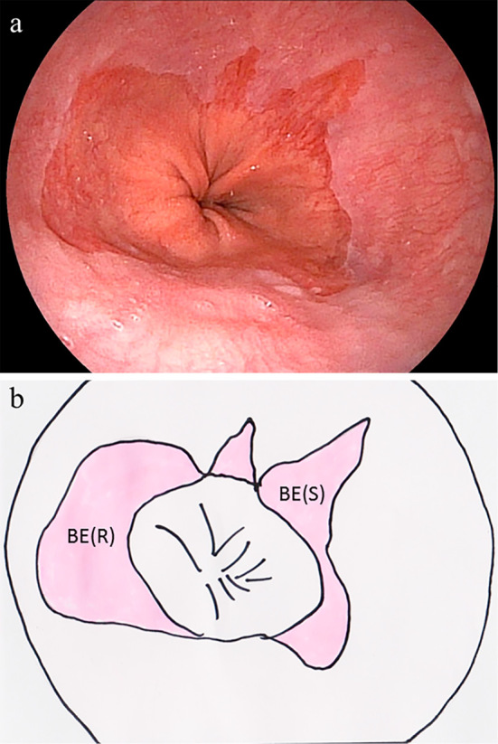
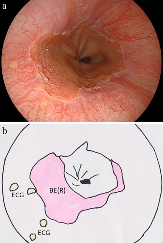
Objective To clarify the relationship between the shape and circumferential location of non-circumferential short-segment Barrett's esophagus (SSBE). Methods We examined 3,788 subjects (2,497 males, 1,291 females; mean age 52.4 years) who underwent upper GI endoscopy as part of a detailed medical checkup. The presence of columnar-appearing mucosa ≥10 mm long in the distal esophagus was diagnosed as BE and then divided into circumferential and non-circumferential localized types. Localized SSBE was further divided into round and sharp types based on the shape of the proximal margin. Results SSBE was endoscopically observed in 197 subjects (5.2%). The numbers of patients with circumferential SSBE, round localized SSBE, and sharp localized SSBE were 38, 114 and 69, respectively. Round and sharp types of localized SSBE were simultaneously observed in 25 patients. Reflux esophagitis was more frequently observed in subjects with BE, regardless of type, in comparison to those without BE. Round localized SSBE was found mainly in the left posterior wall of the esophagus in a location similar to the main area of the esophageal cardiac glands. In contrast, sharp localized SSBE was observed mainly in the right anterior wall of the esophagus in a location similar to that of esophageal mucosal injury caused by mild type reflux esophagitis. Conclusion The location differs between round and sharp localized SSBE, possibly due to differences in the process of BE development.
目的 阐明非环周性短节段Barrett食管(SSBE)的形态与环周位置之间的关系。方法 我们检查了3788名接受上消化道内镜检查作为详细体检一部分的受试者(男性2497名,女性1291名;平均年龄52.4岁)。将食管远端出现长度≥10 mm的柱状黏膜诊断为BE,然后分为环周型和非环周局限型。局限型SSBE根据近端边缘的形状进一步分为圆形和尖锐型。结果 197名受试者(5.2%)经内镜观察到SSBE。环周型SSBE、圆形局限型SSBE和尖锐局限型SSBE的患者人数分别为38、114和69。25名患者同时观察到圆形和尖锐型局限型SSBE。与无BE的受试者相比,无论类型如何,BE受试者中反流性食管炎的观察频率更高。圆形局限型SSBE主要位于食管左后壁,位置与食管贲门腺的主要区域相似。相比之下,尖锐局限型SSBE主要观察到位于食管右前壁,位置与轻度反流性食管炎引起的食管黏膜损伤部位相似。结论 圆形和尖锐局限型SSBE的位置不同,可能是由于BE发展过程中的差异。