Casdorff Kirstin, Keplinger Tobias, Burgert Ingo
Wood Materials Science, Institute for Building Materials, ETH Zürich, Stefano-Franscini-Platz 3, 8093 Zurich, Switzerland.
Applied Wood Materials, Empa-Swiss Federal Laboratories for Materials Science and Technology, Überlandstrasse 129, 8600 Dübendorf, Switzerland.
Plant Methods. 2017 Jul 25;13:60. doi: 10.1186/s13007-017-0211-5. eCollection 2017.
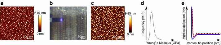
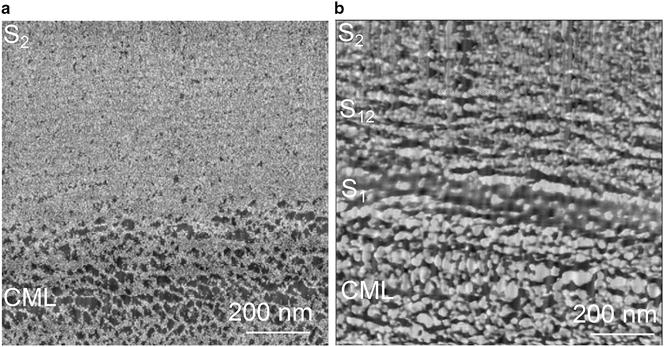
Understanding the arrangement and mechanical properties of wood polymers within the plant cell wall is the basis for unravelling its underlying structure-property relationships. As state of the art Atomic Force Microscopy (AFM) has been used to visualize cell wall layers in contact resonance- and amplitude controlled mode (AC) on embedded samples. Most of the studies have focused on the structural arrangement of the S layer and its lamellar structure.
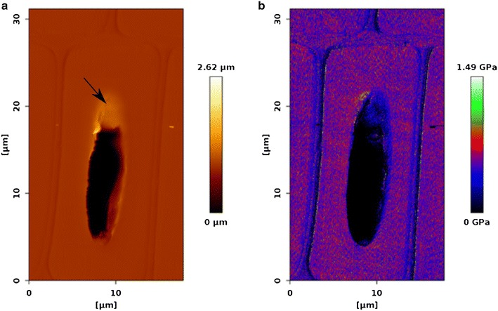
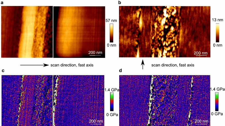
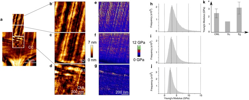
In this work, a protocol for AFM is proposed to characterize the entire cell wall mechanically by quantitative imaging (QI™) at the nanometer level, without embedding the samples. It is shown that the applied protocol allows for distinguishing between the cell wall layers of the compound middle lamella, S, and S of spruce wood based on their Young's Moduli. In the transition zone, S, a stiffness gradient is measured.
The QI™ mode pushes the limit of resolution for mechanical characterization of the plant cell wall to the nanometer range. Comparing QI™- against AC images reveals that the mode of operation strongly influences the visualization of the cell wall.
了解植物细胞壁内木质聚合物的排列和力学性能是揭示其潜在结构-性能关系的基础。作为现有技术,原子力显微镜(AFM)已被用于在嵌入式样品上以接触共振和振幅控制模式(AC)可视化细胞壁层。大多数研究都集中在S层的结构排列及其层状结构上。
在这项工作中,提出了一种AFM方案,通过纳米级的定量成像(QI™)对整个细胞壁进行力学表征,而无需对样品进行包埋。结果表明,所应用的方案能够根据云杉木复合胞间层、S层和S层的杨氏模量区分它们的细胞壁层。在过渡区S层,测量到了刚度梯度。
QI™模式将植物细胞壁力学表征的分辨率极限推至纳米范围。将QI™图像与AC图像进行比较表明,操作模式对细胞壁的可视化有很大影响。