Macfelda Karin, Kapeller Barbara, Holly Alexander, Podesser Bruno K, Losert Udo, Brandes Kersten, Goettel Peter, Mueller Johannes
Department of Biomedical Research (Cell Biology), Medical University of Vienna, Waehringer Guertel 18-20, 1090, Vienna, Austria.
Berlin Heals, Knesebeckstrasse 59-61, 10719, Berlin, Germany.
ESC Heart Fail. 2017 Aug;4(3):291-300. doi: 10.1002/ehf2.12169. Epub 2017 Jun 30.
Beyond the influence of stimulating devices on cardiac excitation, their use in treating patients with heart failure has positive effects on the myocardium at the molecular level. Electrical signals can induce a wide spectrum of effects in living tissue. Therefore, we sought to determine whether applying electrical microcurrent directly to failing hearts leads to functional improvement.
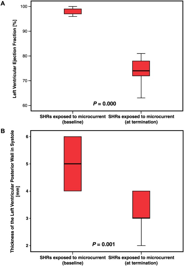
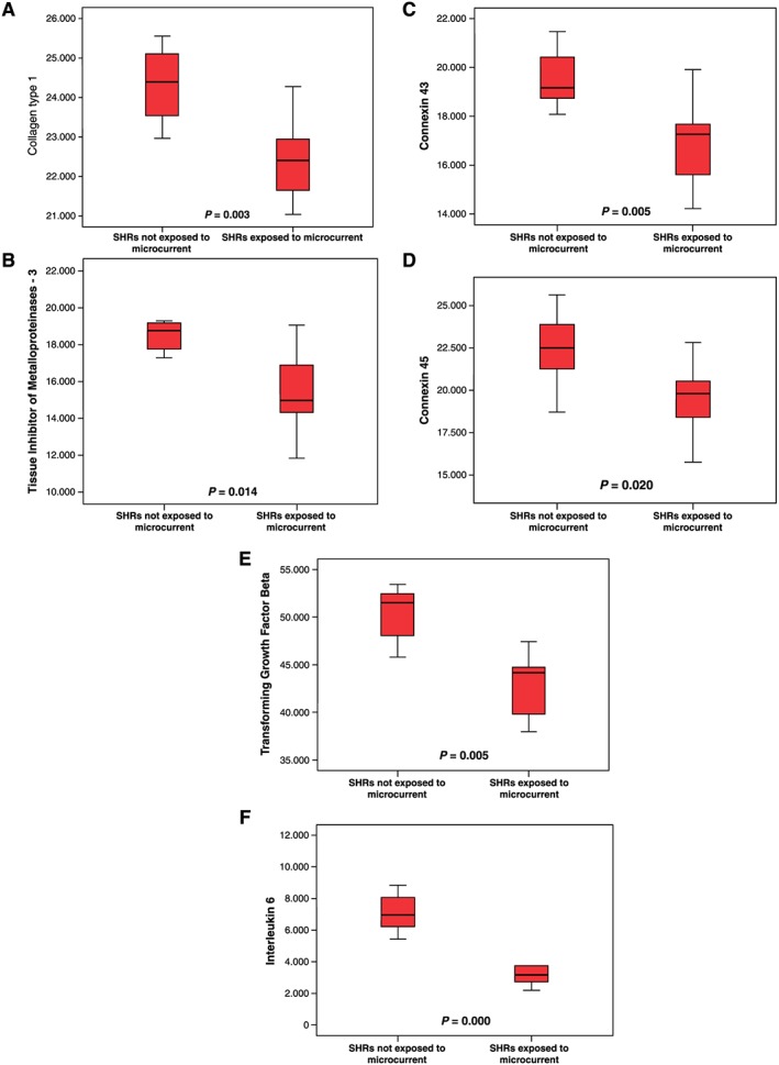
Sixteen male spontaneously hypertensive rats (SHRs) with heart failure underwent application of a patch electrode to the left ventricular epicardium and placement of a subcutaneous counter electrode. The electrode delivered a 0.35 μA microcurrent to nine of the SHRs for 45 ± 3 days; the other seven SHRs were used as controls. At baseline and before the SHRs were humanely put to death, we measured the left ventricular ejection fraction (LVEF) and the thickness of the LV posterior wall during systole and diastole (LVPWs/d). We used quantitative PCR to determine extracellular matrix parameters [collagen I-III, matrix metalloproteinase (MMP)-2, MMP-9, tissue inhibitor of metalloproteinases 3 (TIMP3), TIMP4, connexins (Cxs) 40/43/45, transforming growth factor (TGF)-β, and interleukin (IL)-6]. Among SHRs undergoing microcurrent application, LVEF normalized (mean decrease, 22.8%; P = 0.009), and LVPWs decreased (mean, 35.3%; P = 0.001). Compared with the control group, the SHRs receiving microcurrent exhibited a mean decrease in the gene expression of collagen I (10.6%, P = 0.003), TIMP3 (18.5%, P = 0.005), Cx43 (14.3%, P = 0.003), Cx45 (12.7%, P = 0.020), TGF-β (13.0%, P = 0.005), and IL-6 (53.7%, P = 0.000). Microcurrent application induced no changes in the expression of collagen III, MMP-2, MMP-9, TIMP4, or Cx40.
Applying microcurrent to the LV epicardium of SHRs leads to statistically significant functional improvement and alterations in the levels of inflammatory and extracellular matrix components.
除了刺激装置对心脏兴奋的影响外,其在治疗心力衰竭患者中的应用在分子水平上对心肌有积极作用。电信号可在活组织中诱导多种效应。因此,我们试图确定直接对衰竭心脏施加微电流是否能改善心脏功能。
16只患有心力衰竭的雄性自发性高血压大鼠(SHR)接受了将贴片电极应用于左心室心外膜并放置皮下对电极的操作。电极向9只SHR输送0.35 μA的微电流,持续45±3天;另外7只SHR作为对照。在基线时以及在对SHR实施安乐死之前,我们测量了左心室射血分数(LVEF)以及收缩期和舒张期左心室后壁厚度(LVPWs/d)。我们使用定量PCR来确定细胞外基质参数[I - III型胶原蛋白、基质金属蛋白酶(MMP)-2、MMP - 9、金属蛋白酶组织抑制剂3(TIMP3)、TIMP4、连接蛋白(Cxs)40/43/45、转化生长因子(TGF)-β和白细胞介素(IL)-6]。在接受微电流治疗的SHR中,LVEF恢复正常(平均下降22.8%;P = 0.009),LVPWs下降(平均下降35.3%;P = 0.001)。与对照组相比,接受微电流治疗的SHR在I型胶原蛋白(下降10.6%,P = 0.003)、TIMP3(下降18.5%,P = 0.005)、Cx43(下降14.3%,P = 0.003)、Cx45(下降12.7%,P = 0.020)、TGF -β(下降13.0%,P = 0.005)和IL - 6(下降53.7%,P = 0.000)的基因表达上出现了平均下降。微电流施加未引起III型胶原蛋白、MMP - 2、MMP - 9、TIMP4或Cx40表达的变化。
对SHR的左心室心外膜施加微电流可导致具有统计学意义的功能改善以及炎症和细胞外基质成分水平的改变。