Thao Le Thi Song, Dang Trinh Trung Tri, Khanitchaidecha Wilawan, Channei Duangdao, Nakaruk Auppatham
Department of Civil Engineering, Faculty of Engineering, Naresuan University, Phitsanulok 65000, Thailand.
Centre of Excellence for Innovation and Technology for Water Treatment, Naresuan University, Phitsanulok 65000, Thailand.
Materials (Basel). 2017 Jan 30;10(2):122. doi: 10.3390/ma10020122.
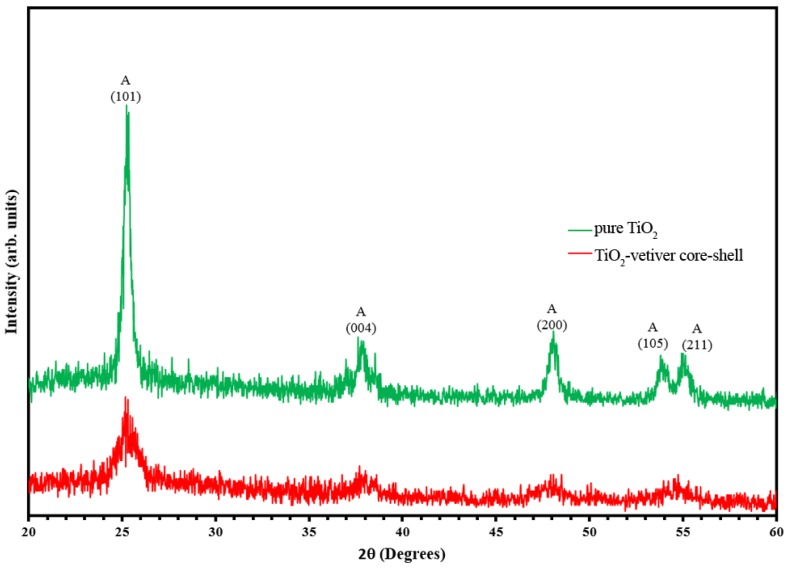
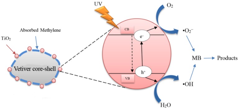
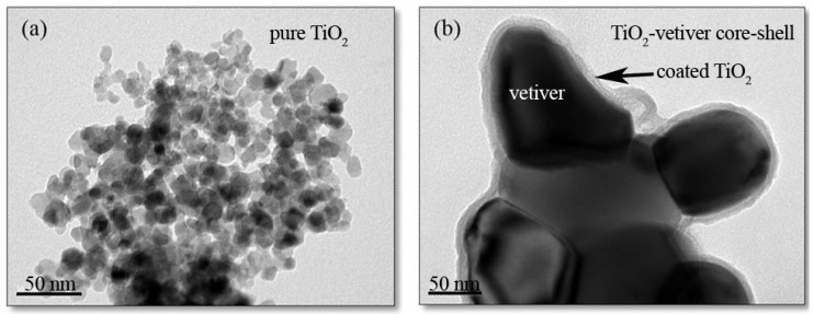
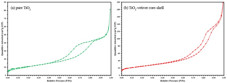
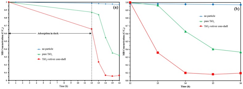
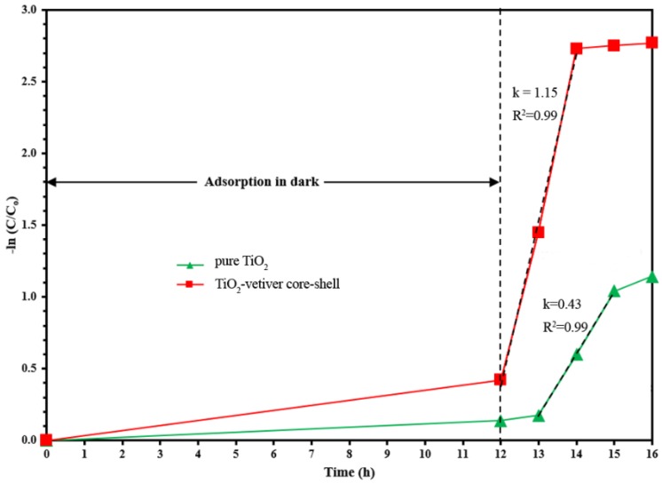
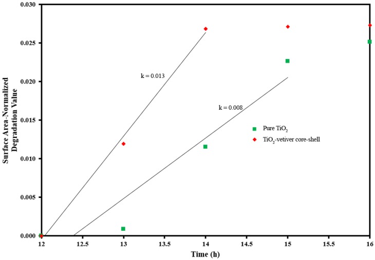
The properties and photocatalytic performance of anatase nanoparticles of pure TiO₂ and a core-shell structure of TiO₂ on calcined vetiver grass leaves have been compared. Samples were fabricated by sol-gel and heating at 450 °C for 5h.The comparison was based on data for X-ray diffraction(XRD), UV-Vis spectrophotometry, photoluminescence, transmission electron microscopy, specific surface area measurement, pore volume assessment, and methylene blue degradation testing. The results showed that the pure TiO2 consisted of agglomerated equiaxed nanoparticles of individual grain sizes in the range 10-20 nm. In contrast, the TiO₂-vetiver composite exhibited a core-shell structure consisting of a carbonaceous core and TiO₂ shell of thickness 10-15nm. These features influenced the photocatalytic performance in such a way that the lower crosssectional area, greater surface area, and higher pore volume of the TiO₂ shell increased the number of active sites, reduced the charge carrier diffusion distance, and reduced the recombination rate, thereby improving the photocatalytic activity. This improvement derived from morphological characteristics rather than crystallographic, semiconducting, or optical properties. The improved performance of the TiO₂-vetiver core-shell was unexpected since the X-ray diffraction data showed that the crystallinity of the TiO₂ was lower than that of the pure TiO₂. These outcomes are attributed to the reducing effect of the carbon on the TiO₂ during heating, thereby facilitating the formation of oxygen vacancies, which enhance charge separation and hence photocatalysis by TiO₂.
已对纯TiO₂锐钛矿型纳米颗粒以及煅烧香根草叶片上TiO₂核壳结构的性质和光催化性能进行了比较。通过溶胶-凝胶法制备样品,并在450℃下加热5小时。该比较基于X射线衍射(XRD)、紫外-可见分光光度法、光致发光、透射电子显微镜、比表面积测量、孔体积评估和亚甲基蓝降解测试的数据。结果表明,纯TiO₂由粒径在10-20nm范围内的团聚等轴纳米颗粒组成。相比之下,TiO₂-香根草复合材料呈现出核壳结构,由碳质核和厚度为10-15nm的TiO₂壳组成。这些特征以如下方式影响光催化性能:TiO₂壳较低的横截面积、较大的表面积和较高的孔体积增加了活性位点的数量,减小了电荷载流子的扩散距离,并降低了复合率,从而提高了光催化活性。这种改善源于形态特征而非晶体学、半导体或光学性质。TiO₂-香根草核壳的性能改善出乎意料,因为X射线衍射数据表明TiO₂的结晶度低于纯TiO₂。这些结果归因于加热过程中碳对TiO₂的还原作用,从而促进了氧空位的形成,增强了电荷分离,进而提高了TiO₂的光催化性能。