• 文献检索
  • 文档翻译
  • 深度研究
  • 学术资讯
  • Suppr Zotero 插件Zotero 插件
  • 邀请有礼
  • 套餐&价格
  • 历史记录
应用&插件
Suppr Zotero 插件Zotero 插件浏览器插件Mac 客户端Windows 客户端微信小程序
定价
高级版会员购买积分包购买API积分包
服务
文献检索文档翻译深度研究API 文档MCP 服务
关于我们
关于 Suppr公司介绍联系我们用户协议隐私条款
关注我们

Suppr 超能文献

核心技术专利:CN118964589B侵权必究
粤ICP备2023148730 号-1Suppr @ 2026

文献检索

告别复杂PubMed语法,用中文像聊天一样搜索,搜遍4000万医学文献。AI智能推荐,让科研检索更轻松。

立即免费搜索

文件翻译

保留排版,准确专业,支持PDF/Word/PPT等文件格式,支持 12+语言互译。

免费翻译文档

深度研究

AI帮你快速写综述,25分钟生成高质量综述,智能提取关键信息,辅助科研写作。

立即免费体验

慢性给予和撤去钒后小鼠脑内金属分布及神经炎症特征

Brain Metal Distribution and Neuro-Inflammatory Profiles after Chronic Vanadium Administration and Withdrawal in Mice.

作者信息

Folarin Oluwabusayo R, Snyder Amanda M, Peters Douglas G, Olopade Funmilayo, Connor James R, Olopade James O

机构信息

Department of Medical Laboratory Science, Ladoke Akintola University of TechnologyOsogbo, Nigeria.

Department of Neurosurgery, Pennsylvania State College of MedicineHershey, PA, United States.

出版信息

Front Neuroanat. 2017 Jul 25;11:58. doi: 10.3389/fnana.2017.00058. eCollection 2017.

DOI:10.3389/fnana.2017.00058
PMID:28790895
原文链接:https://pmc.ncbi.nlm.nih.gov/articles/PMC5524677/
Abstract

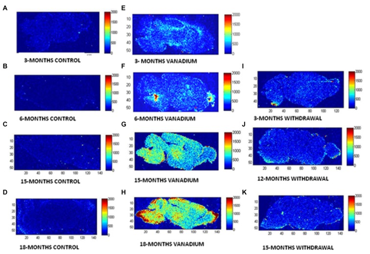
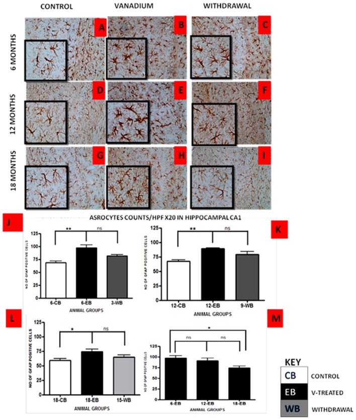
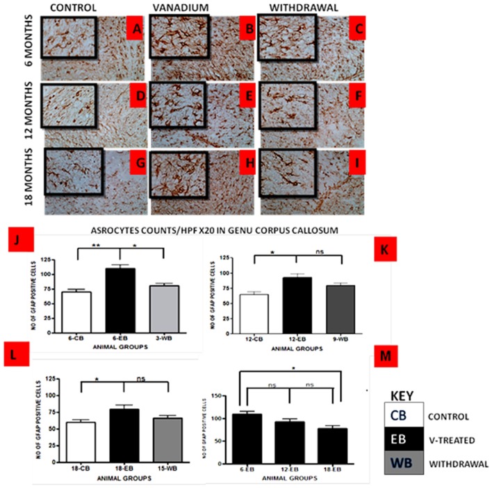
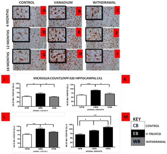
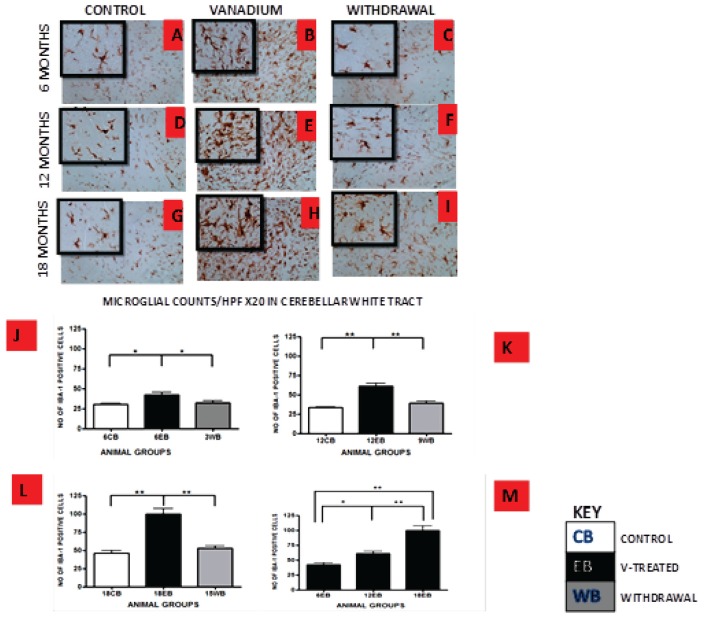
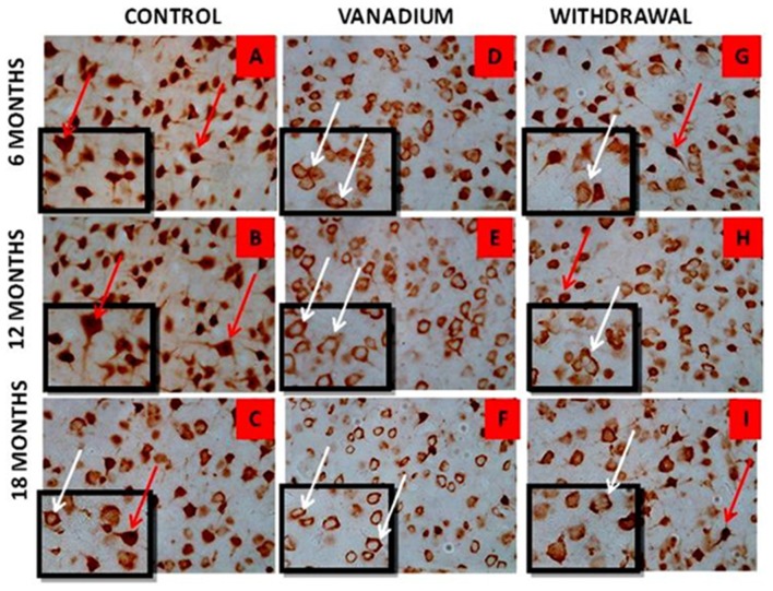
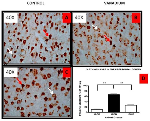
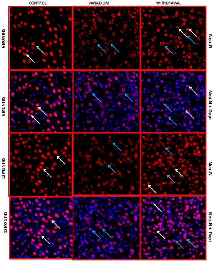
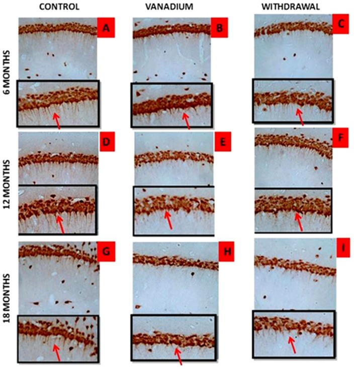
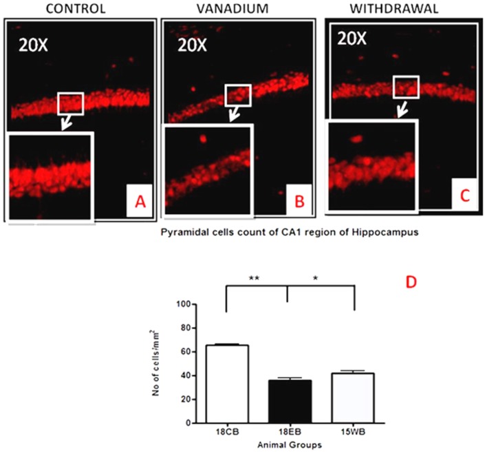
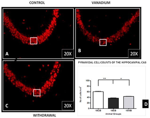
Vanadium is a potentially toxic environmental pollutant and induces oxidative damage in biological systems including the central nervous system (CNS). Its deposition in brain tissue may be involved in the pathogenesis of certain neurological disorders which after prolonged exposure can culminate into more severe pathology. Most studies on vanadium neurotoxicity have been done after acute exposure but in reality some populations are exposed for a lifetime. This work was designed to ascertain neurodegenerative consequences of chronic vanadium administration and to investigate the progressive changes in the brain after withdrawal from vanadium treatment. A total of 85 male BALB/c mice were used for the experiment and divided into three major groups of vanadium treated (intraperitoneally (i.p.) injected with 3 mg/kg body weight of sodium metavanadate and sacrificed every 3 months till 18 months); matched controls; and animals that were exposed to vanadium for 3 months and thereafter the metal was withdrawn. Brain tissues were obtained after animal sacrifice. Sagittal cut sections of paraffin embedded tissue (5 μm) were analyzed by the Laser ablation-inductively coupled plasma-mass spectrometry (LA-ICP-MS) to show the absorption and distribution of vanadium metal. Also, Haematoxylin and Eosin (H&E) staining of brain sections, and immunohistochemistry for Microglia (Iba-1), Astrocytes (GFAP), Neurons (Neu-N) and Neu-N + 4',6-diamidine-2'-pheynylindole dihydrochloride (Dapi) Immunofluorescent labeling were observed for morphological and morphometric parameters. The LA-ICP-MS results showed progressive increase in vanadium uptake with time in different brain regions with prediction for regions like the olfactory bulb, brain stem and cerebellum. The withdrawal brains still show presence of vanadium metal in the brain slightly more than the controls. There were morphological alterations (of the layering profile, nuclear shrinkage) in the prefrontal cortex, cellular degeneration (loss of dendritic arborization) and cell death in the Hippocampal CA1 pyramidal cells and Purkinje cells of the cerebellum, including astrocytic and microglial activation in vanadium exposed brains which were all attenuated in the withdrawal group. With exposure into old age, the evident neuropathology was microgliosis, while progressive astrogliosis became more attenuated. We have shown that chronic administration of vanadium over a lifetime in mice resulted in metal accumulation which showed regional variabilities with time. The metal profile and pathological effects were not completely eliminated from the brain even after a long time withdrawal from vanadium metal.

摘要

钒是一种具有潜在毒性的环境污染物,可在包括中枢神经系统(CNS)在内的生物系统中引发氧化损伤。其在脑组织中的沉积可能参与某些神经系统疾病的发病机制,长期接触后可能会发展为更严重的病理状况。大多数关于钒神经毒性的研究都是在急性暴露后进行的,但实际上有些人群会终身接触钒。这项研究旨在确定长期给予钒后的神经退行性后果,并研究停止钒治疗后大脑的渐进性变化。总共85只雄性BALB/c小鼠用于该实验,分为三大组:钒处理组(腹腔注射3mg/kg体重的偏钒酸钠,每3个月处死一批,直至18个月);匹配对照组;以及接触钒3个月后停止接触的动物组。动物处死后获取脑组织。对石蜡包埋组织的矢状切片(5μm)进行激光烧蚀-电感耦合等离子体质谱(LA-ICP-MS)分析,以显示钒金属的吸收和分布情况。此外,对脑切片进行苏木精和伊红(H&E)染色,并对小胶质细胞(Iba-1)、星形胶质细胞(GFAP)、神经元(Neu-N)以及神经元+4',6-二脒基-2'-苯基吲哚二盐酸盐(Dapi)进行免疫荧光标记,以观察形态学和形态计量学参数。LA-ICP-MS结果显示,随着时间的推移,不同脑区的钒摄取量逐渐增加,嗅球、脑干和小脑等区域的摄取量预计会更高。停止接触钒的小鼠大脑中钒金属的含量仍略高于对照组。在钒暴露组的大脑中,前额叶皮质出现了形态学改变(分层结构、核收缩)、细胞变性(树突分支减少)以及海马CA1锥体细胞和小脑浦肯野细胞的细胞死亡,包括星形胶质细胞和小胶质细胞的激活,而在停止接触钒的组中这些变化均有所减轻。随着年龄增长,明显的神经病理学变化是小胶质细胞增生,而进行性星形胶质细胞增生则有所减轻。我们已经表明,小鼠终身长期给予钒会导致金属蓄积,且金属蓄积量会随时间呈现区域差异。即使在停止接触钒金属很长时间后,大脑中的金属分布和病理效应也并未完全消除。

https://cdn.ncbi.nlm.nih.gov/pmc/blobs/405d/5524677/40288836bf14/fnana-11-00058-g0012.jpg
https://cdn.ncbi.nlm.nih.gov/pmc/blobs/405d/5524677/0cf531b9b82d/fnana-11-00058-g0001.jpg
https://cdn.ncbi.nlm.nih.gov/pmc/blobs/405d/5524677/e587749de016/fnana-11-00058-g0002.jpg
https://cdn.ncbi.nlm.nih.gov/pmc/blobs/405d/5524677/0eaac5890568/fnana-11-00058-g0003.jpg
https://cdn.ncbi.nlm.nih.gov/pmc/blobs/405d/5524677/ec5317a289b6/fnana-11-00058-g0004.jpg
https://cdn.ncbi.nlm.nih.gov/pmc/blobs/405d/5524677/bd001869a33e/fnana-11-00058-g0005.jpg
https://cdn.ncbi.nlm.nih.gov/pmc/blobs/405d/5524677/c21941b11ccb/fnana-11-00058-g0006.jpg
https://cdn.ncbi.nlm.nih.gov/pmc/blobs/405d/5524677/69087f62f2e6/fnana-11-00058-g0007.jpg
https://cdn.ncbi.nlm.nih.gov/pmc/blobs/405d/5524677/d6f0bf6a8222/fnana-11-00058-g0008.jpg
https://cdn.ncbi.nlm.nih.gov/pmc/blobs/405d/5524677/71ba987c03c6/fnana-11-00058-g0009.jpg
https://cdn.ncbi.nlm.nih.gov/pmc/blobs/405d/5524677/df8199c7282a/fnana-11-00058-g0010.jpg
https://cdn.ncbi.nlm.nih.gov/pmc/blobs/405d/5524677/e18b48eb89d1/fnana-11-00058-g0011.jpg
https://cdn.ncbi.nlm.nih.gov/pmc/blobs/405d/5524677/40288836bf14/fnana-11-00058-g0012.jpg

相似文献

1
Brain Metal Distribution and Neuro-Inflammatory Profiles after Chronic Vanadium Administration and Withdrawal in Mice.慢性给予和撤去钒后小鼠脑内金属分布及神经炎症特征
Front Neuroanat. 2017 Jul 25;11:58. doi: 10.3389/fnana.2017.00058. eCollection 2017.
2
Novel NMDA-receptor antagonists ameliorate vanadium neurotoxicity.新型N-甲基-D-天冬氨酸受体拮抗剂可改善钒神经毒性。
Naunyn Schmiedebergs Arch Pharmacol. 2020 Sep;393(9):1729-1738. doi: 10.1007/s00210-020-01882-6. Epub 2020 May 9.
3
Changes in the brain antioxidant profile after chronic vanadium administration in mice.慢性钒染毒后小鼠脑抗氧化剂谱的变化。
Metab Brain Dis. 2018 Apr;33(2):377-385. doi: 10.1007/s11011-017-0070-9. Epub 2017 Jul 25.
4
Cerebellar and Olfactory Bulb Perturbations Induced by Vanadium Neurotoxicity in the African Giant Rat (Cricetomys gambianus, Waterhouse).钒神经毒性对非洲巨鼠(冈比亚囊鼠,沃特豪斯)小脑和嗅球的影响
Niger J Physiol Sci. 2023 Dec 31;38(2):135-143. doi: 10.54548/njps.v38i2.3.
5
The ameliorative effects of a phenolic derivative of leave against vanadium-induced neurotoxicity in mice.一种树叶酚类衍生物对钒诱导的小鼠神经毒性的改善作用。
IBRO Rep. 2020 Jul 12;9:164-182. doi: 10.1016/j.ibror.2020.07.004. eCollection 2020 Dec.
6
Memory Deficit Recovery after Chronic Vanadium Exposure in Mice.慢性钒暴露后小鼠记忆缺陷的恢复。
Oxid Med Cell Longev. 2016;2016:4860582. doi: 10.1155/2016/4860582. Epub 2016 Jan 19.
7
DNA damage in nasal and brain tissues of canines exposed to air pollutants is associated with evidence of chronic brain inflammation and neurodegeneration.暴露于空气污染物的犬类鼻腔和脑组织中的DNA损伤与慢性脑部炎症和神经退行性变的证据有关。
Toxicol Pathol. 2003 Sep-Oct;31(5):524-38. doi: 10.1080/01926230390226645.
8
Vanadium (V)-induced neurotoxicity in the rat central nervous system: a histo-immunohistochemical study.钒(V)诱导大鼠中枢神经系统神经毒性的组织免疫组化研究
Drug Chem Toxicol. 2005;28(3):329-44. doi: 10.1081/dct-200064496.
9
Regional Myelin and Axon Damage and Neuroinflammation in the Adult Mouse Brain After Long-Term Postnatal Vanadium Exposure.长期产后钒暴露后成年小鼠大脑中的局部髓鞘和轴突损伤及神经炎症
J Neuropathol Exp Neurol. 2016 Sep;75(9):843-54. doi: 10.1093/jnen/nlw058. Epub 2016 Jul 6.
10
Neurobehavioural and Histological Study of the Effects of Low-Dose and High-Dose Vanadium in Brain, Liver and Kidney of Mice.神经行为和组织学研究低剂量和高剂量钒对小鼠脑、肝和肾的影响。
Niger J Physiol Sci. 2023 May 30;38(1):47-56. doi: 10.54548/njps.v38i1.8.

引用本文的文献

1
Chronic Vanadium Exposure Promotes Aggregation of Alpha-Synuclein, Tau and Amyloid Beta in Mouse Brain.长期接触钒会促进小鼠大脑中α-突触核蛋白、tau蛋白和β-淀粉样蛋白的聚集。
J Neurochem. 2025 May;169(5):e70082. doi: 10.1111/jnc.70082.
2
Arsenic and vanadium co-exposure induced cerebellar neurotoxicity: aggravates apoptosis, inflammation, oxidative stress, and locomotor deficits in juvenile BALB/c mice.砷和钒共同暴露诱导小脑神经毒性:加重幼年BALB/c小鼠的细胞凋亡、炎症、氧化应激和运动功能障碍。
Anat Cell Biol. 2025 Jun 30;58(2):229-246. doi: 10.5115/acb.24.116. Epub 2025 Mar 26.
3
ZA-II-05, a novel NMDA-receptor antagonist reverses vanadium-induced neurotoxicity in Caenorhabditis elegans (C. elegans).

本文引用的文献

1
The Deterioration Seen in Myelin Related Morphophysiology in Vanadium Exposed Rats is Partially Protected by Concurrent Iron Deficiency.钒暴露大鼠髓鞘相关形态生理学的恶化部分受到同时缺铁的保护。
Niger J Physiol Sci. 2016 Aug 30;31(1):11-22.
2
Regional Myelin and Axon Damage and Neuroinflammation in the Adult Mouse Brain After Long-Term Postnatal Vanadium Exposure.长期产后钒暴露后成年小鼠大脑中的局部髓鞘和轴突损伤及神经炎症
J Neuropathol Exp Neurol. 2016 Sep;75(9):843-54. doi: 10.1093/jnen/nlw058. Epub 2016 Jul 6.
3
Memory Deficit Recovery after Chronic Vanadium Exposure in Mice.
ZA-II-05,一种新型 NMDA 受体拮抗剂,可逆转秀丽隐杆线虫(C. elegans)中钒诱导的神经毒性。
BMC Neurosci. 2024 Oct 28;25(1):56. doi: 10.1186/s12868-024-00902-y.
4
Exploring the Biological Effects of Anti-Diabetic Vanadium Compounds in the Liver, Heart and Brain.探索抗糖尿病钒化合物对肝脏、心脏和大脑的生物学效应。
Diabetes Metab Syndr Obes. 2024 Sep 4;17:3267-3278. doi: 10.2147/DMSO.S417700. eCollection 2024.
5
Attenuation of Vanadium-Induced Neurotoxicity in Rat Hippocampal Slices (In Vitro) and Mice (In Vivo) by ZA-II-05, a Novel NMDA-Receptor Antagonist.新型 NMDA 受体拮抗剂 ZA-II-05 对大鼠海马脑片(体外)和小鼠(体内)钒诱导的神经毒性的衰减作用。
Int J Mol Sci. 2023 Nov 24;24(23):16710. doi: 10.3390/ijms242316710.
6
Flavonoid glycoside fraction of extract modulates antioxidants imbalance in vanadium-induced brain damage.提取物中的黄酮糖苷部分可调节钒诱导的脑损伤中的抗氧化剂失衡。
AIMS Neurosci. 2023 Jun 29;10(2):178-189. doi: 10.3934/Neuroscience.2023015. eCollection 2023.
7
Disease-associated metabolic pathways affected by heavy metals and metalloid.受重金属和类金属影响的疾病相关代谢途径。
Toxicol Rep. 2023 Apr 24;10:554-570. doi: 10.1016/j.toxrep.2023.04.010. eCollection 2023.
8
Biological Consequences of Vanadium Effects on Formation of Reactive Oxygen Species and Lipid Peroxidation.钒对活性氧形成和脂质过氧化的生物学影响。
Int J Mol Sci. 2023 Mar 11;24(6):5382. doi: 10.3390/ijms24065382.
9
Vanadium improves memory and spatial learning and protects the pyramidal cells of the hippocampus in juvenile hydrocephalic mice.钒可改善幼年脑积水小鼠的记忆力和空间学习能力,并保护海马体的锥体细胞。
Front Neurol. 2023 Feb 9;14:1116727. doi: 10.3389/fneur.2023.1116727. eCollection 2023.
10
Impact of Environmental Risk Factors on Mitochondrial Dysfunction, Neuroinflammation, Protein Misfolding, and Oxidative Stress in the Etiopathogenesis of Parkinson's Disease.环境风险因素对帕金森病发病机制中线粒体功能障碍、神经炎症、蛋白质错误折叠和氧化应激的影响。
Int J Mol Sci. 2022 Sep 16;23(18):10808. doi: 10.3390/ijms231810808.
慢性钒暴露后小鼠记忆缺陷的恢复。
Oxid Med Cell Longev. 2016;2016:4860582. doi: 10.1155/2016/4860582. Epub 2016 Jan 19.
4
Morphological plasticity of astroglia: Understanding synaptic microenvironment.星形胶质细胞的形态可塑性:理解突触微环境。
Glia. 2015 Dec;63(12):2133-51. doi: 10.1002/glia.22821. Epub 2015 Mar 18.
5
Neurobehavioral and cytotoxic effects of vanadium during oligodendrocyte maturation: a protective role for erythropoietin.钒在少突胶质细胞成熟过程中的神经行为和细胞毒性作用:促红细胞生成素的保护作用。
Environ Toxicol Pharmacol. 2014 Jul;38(1):98-111. doi: 10.1016/j.etap.2014.05.001. Epub 2014 May 10.
6
Taurine improves the spatial learning and memory ability impaired by sub-chronic manganese exposure.牛磺酸可改善亚慢性锰暴露所致的空间学习和记忆能力损害。
J Biomed Sci. 2014 May 24;21(1):51. doi: 10.1186/1423-0127-21-51.
7
Vanadium exposure induces olfactory dysfunction in an animal model of metal neurotoxicity.钒暴露在金属神经毒性动物模型中诱发嗅觉功能障碍。
Neurotoxicology. 2014 Jul;43:73-81. doi: 10.1016/j.neuro.2013.12.004. Epub 2013 Dec 18.
8
Vanadium exposure-induced neurobehavioral alterations among Chinese workers.钒接触致中国作业工人神经行为改变。
Neurotoxicology. 2013 May;36:49-54. doi: 10.1016/j.neuro.2013.02.008. Epub 2013 Mar 7.
9
Three-dimensional atlas of iron, copper, and zinc in the mouse cerebrum and brainstem.鼠脑大脑皮质和脑干中铁、铜、锌的三维图谱。
Anal Chem. 2012 May 1;84(9):3990-7. doi: 10.1021/ac300374x. Epub 2012 Apr 10.
10
Vertical administration of vanadium through lactation induces behavioural and neuromorphological changes: protective role of vitamin E.通过哺乳垂直给予钒会引起行为和神经形态学变化:维生素E的保护作用。
Niger J Physiol Sci. 2011 Nov 23;26(1):55-60.