Department of Anesthesiology and Intensive Care Unit, Eastern Hepatobiliary Surgery Hospital, Second Military Medical University, Shanghai 200438, P.R. China.
Department of Anesthesiology, 309th hospital of CPLA, Beijing 100091, P.R. China.
Mol Med Rep. 2017 Oct;16(4):4429-4436. doi: 10.3892/mmr.2017.7173. Epub 2017 Aug 4.
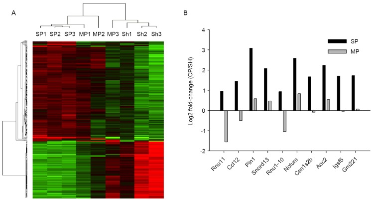
There is emerging evidence on the mechanisms of pancreatic cancer pain. Following the establishment of an orthotropic transplantation model of pancreatic cancer, microarray analysis was performed to identify changes in the expression levels of pain‑associated genes in the spinal cord. A mouse model of pancreatic cancer‑induced pain was established by implanting SW 1990 cells into the pancreases of female BALB/c‑nu mice. The survival rate and body weight were measured following orthotropic transplantation. Gross anatomical techniques and hematoxylin and eosin staining were used to analyze the pancreatic tumor tissue. Multiple behavioral tests were also performed to assess pain‑associated responses. Additionally, using samples from mice with or without observable pain, microarray analysis was performed to determine the gene expression profiles in the spinal cord dorsal horn. The survival rate of mice with pancreatic cancer was high during the initial 3 weeks post‑surgery, although the body weight decreased progressively. Gross anatomical techniques demonstrated that the tumor size increased significantly following the surgery, and this result was confirmed by solid tumor masses in the pancreatic tissues of the mouse model. Observable pain behavioral responses were also examined in the pancreatic cancer model by measuring the mechanical threshold of the abdominal skin, hunching behavior and visceromotor responses. The profiles of 10 pain specific‑associated genes in the spinal cord dorsal horn that accurately reflect the molecular pathological progression of disease were also identified. In conclusion, the present study has developed a novel animal model of pancreatic cancer pain in BALB/c‑nu mice that resembles human pancreatic cancer pain, and the expression of pain‑associated genes in the spinal cord dorsal horn has been profiled. The results of the present study may further the understanding of the molecular mechanisms that mediate pancreatic cancer pain.
胰腺癌疼痛的发生机制方面已有一些新的证据。在建立胰腺癌原位移植模型后,通过基因芯片分析确定脊髓中与疼痛相关基因的表达水平变化。将 SW1990 细胞植入雌性 BALB/c-裸鼠胰腺中,建立胰腺癌诱导痛模型。通过原位移植后测量存活率和体重来评估动物模型。使用大体解剖技术和苏木精-伊红染色分析胰腺肿瘤组织。还进行了多种行为学测试来评估与疼痛相关的反应。此外,使用有或无明显疼痛的小鼠样本进行基因芯片分析,以确定脊髓背角的基因表达谱。在手术后的前 3 周,胰腺癌小鼠的存活率较高,尽管体重逐渐下降。大体解剖技术显示手术后肿瘤大小显著增加,并且在小鼠胰腺组织中的实体瘤块也证实了这一结果。还通过测量腹部皮肤的机械阈值、缩颈行为和内脏运动反应来检查胰腺癌模型中的可观察到的疼痛行为反应。还确定了 10 个脊髓背角中与疼痛特异性相关的基因,这些基因的表达谱准确反映了疾病的分子病理进展。总之,本研究在 BALB/c-裸鼠中建立了一种新的胰腺癌痛动物模型,该模型类似于人类胰腺癌痛,并且对脊髓背角中与疼痛相关基因的表达进行了分析。本研究的结果可能有助于进一步了解介导胰腺癌痛的分子机制。