aDepartment of Radiology bDepartment of Health Sciences Research cDepartment of Information Technology dDepartment of Obstetrics and Gynecology eDepartment of Psychiatry and Psychology fDepartment of Neurology gDivisions of Biomedical Statistics and Informatics and Epidemiology, Department of Health Sciences Research hDepartment of Surgery iDepartment of Physiology and Biomedical Engineering jDivision of Nephrology and Hypertension, Department of Internal Medicine, Mayo Clinic, Rochester, Minnesota, USA.
J Hypertens. 2017 Dec;35(12):2479-2485. doi: 10.1097/HJH.0000000000001492.
Women with a history of preeclampsia are at an increased risk of hypertension and structural brain changes. However, the combined effect of both preeclampsia and late-life hypertension on brain structural changes is not known and was investigated in this study.
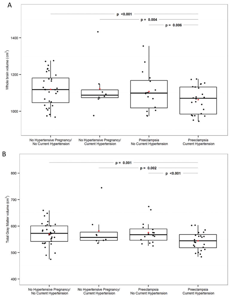
Participants were identified from the population-based Rochester Epidemiology Project cohort. Four groups of women were recruited and investigated in this study: first, women with a history of normotensive pregnancy who have late-life hypertension (n = 8, median age = 62), second, women with a history of normotensive pregnancy who do not have late-life hypertension (n = 32, median age = 59), third, women with a history of preeclampsia who have late-life hypertension (n = 24, median age = 60), and fourth, women with a history of preeclampsia who do not have late-life hypertension (n = 16, median age = 57). Cerebrovascular disease lesions on MRI, and total gray matter volumes were assessed.
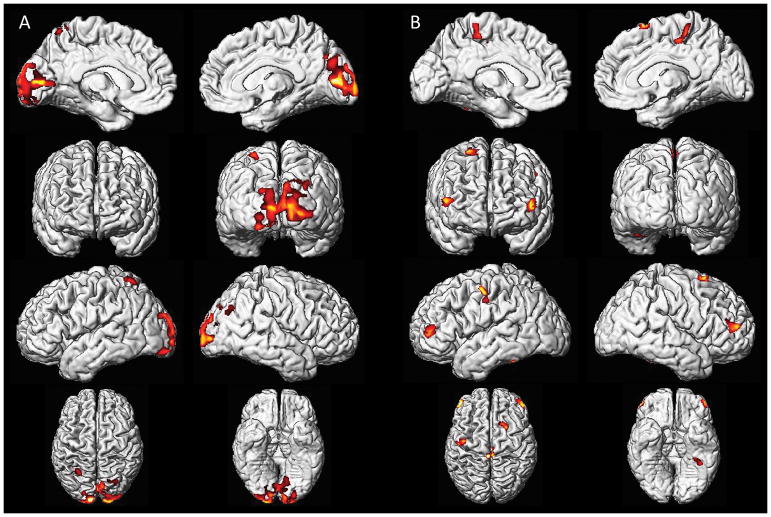
Total gray matter volumes were smaller in women with a history of preeclampsia and late-life hypertension compared with the other groups. Voxel-based morphometry demonstrated that the volume changes were localized to the posterior brain regions, particularly the occipital lobe gray matter in women with a history of preeclampsia and late-life hypertension.
Having late-life hypertension superimposed on a history of preeclampsia affects the brain structure differently than having either a history of preeclampsia alone or a history of normotensive pregnancy either with or without late-life hypertension.
患有先兆子痫的女性患高血压和结构性脑改变的风险增加。然而,先兆子痫和老年期高血压对脑结构变化的综合影响尚不清楚,本研究对此进行了调查。
参与者从基于人群的罗切斯特流行病学项目队列中确定。本研究招募并调查了四组女性:第一组,有正常妊娠史且患有老年期高血压的女性(n=8,中位年龄 62 岁);第二组,有正常妊娠史且无老年期高血压的女性(n=32,中位年龄 59 岁);第三组,有先兆子痫史且患有老年期高血压的女性(n=24,中位年龄 60 岁);第四组,有先兆子痫史且无老年期高血压的女性(n=16,中位年龄 57 岁)。评估 MRI 上的脑血管疾病病变和总灰质体积。
与其他组相比,有先兆子痫和老年期高血压史的女性总灰质体积较小。体素形态计量学显示,体积变化局限于大脑后部区域,尤其是有先兆子痫和老年期高血压史的女性的枕叶灰质。
与单独患有先兆子痫史或正常妊娠史(无论是否伴有老年期高血压)相比,老年期高血压叠加先兆子痫病史对脑结构的影响不同。