Kouwenhoven Willemieke M, von Oerthel Lars, Smidt Marten P
Swammerdam Institute for Life Sciences, University of Amsterdam, Amsterdam, the Netherlands.
PLoS One. 2017 Aug 11;12(8):e0182421. doi: 10.1371/journal.pone.0182421. eCollection 2017.
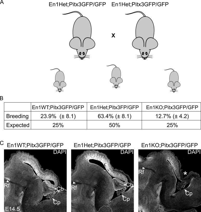
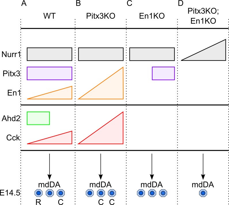
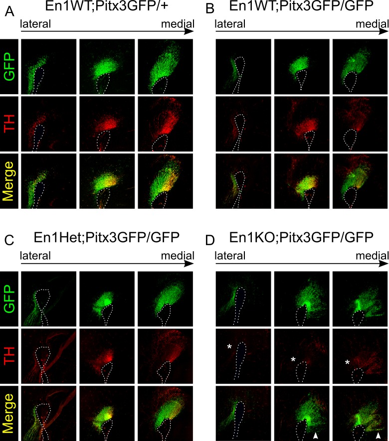
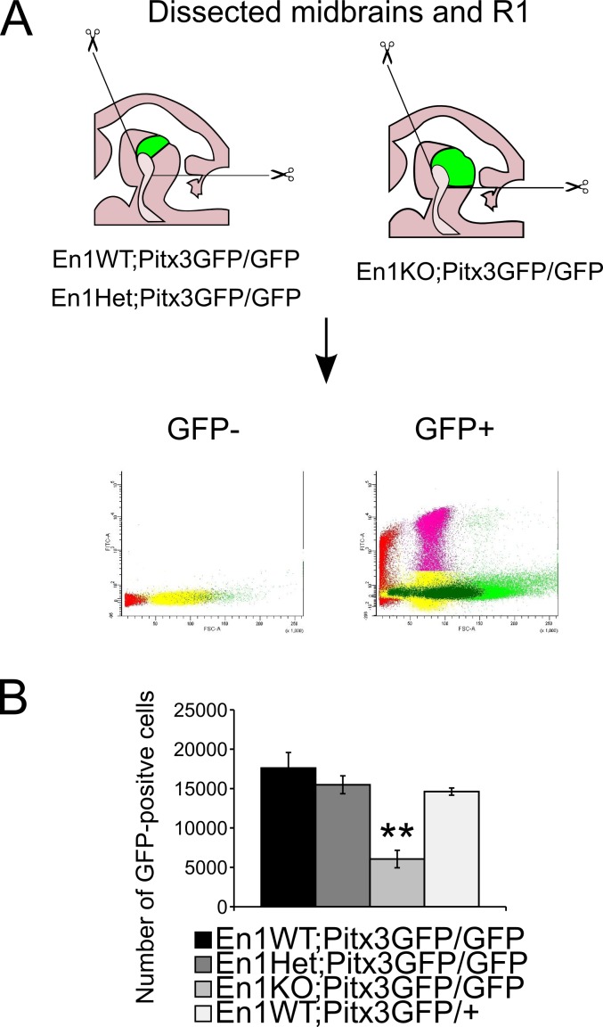
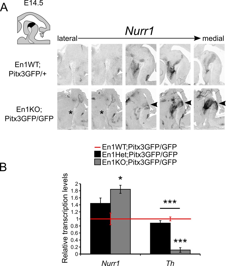
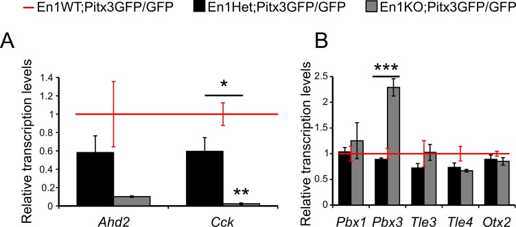
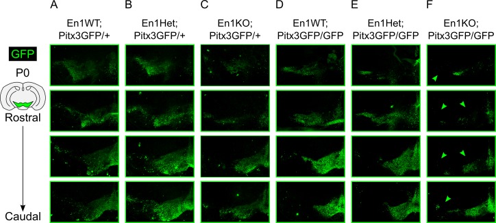
Mesodiencephalic dopaminergic (mdDA) neurons are located in the ventral midbrain. These neurons form the substantia nigra (SNc) and the ventral tegmental area (VTA). Two transcription factors that play important roles in the process of terminal differentiation and subset-specification of mdDA neurons, are paired-like homeodomain transcription factor 3 (Pitx3), and homeobox transcription factor Engrailed 1 (En1). We previously investigated the single Pitx3KO and En1KO and observed important changes in the survival of mdDA neurons of the SNc and VTA as well as altered expression of pivotal rostral- and caudal-markers, Ahd2 and Cck, respectively. To refine our understanding of the regional-specific relationships between En1 and Pitx3 and their (combined) role in the programming mdDA neurons on the rostral-to-caudal axis, we created double En1tm1Alj/tm1Alj;Pitx3gfp/gfp (En1KO;Pitx3GFP/GFP) animals. Here we report, that in absence of En1 and Pitx3, only a limited number of mdDA neurons are present at E14.5. These mdDA neurons have a rudimentary dopaminergic cell fate, as they express Nurr1, Pbx3 and Otx2 but have lost their rostral or caudal subset identity. Furthermore, we report that the expression of Cck depends on En1 expression, while (in contrast) both Pitx3 and En1 are involved in the initiation of Ahd2 expression. Thus we reveal in this manuscript that regulated levels of Pitx3 and En1 control the size and rostral/caudal-identity of the mdDA neuronal population.
中脑多巴胺能(mdDA)神经元位于腹侧中脑。这些神经元形成黑质(SNc)和腹侧被盖区(VTA)。在mdDA神经元的终末分化和亚群特异性过程中发挥重要作用的两个转录因子是配对样同源域转录因子3(Pitx3)和同源框转录因子Engrailed 1(En1)。我们之前研究了单个Pitx3基因敲除和En1基因敲除,并观察到SNc和VTA的mdDA神经元存活情况发生了重要变化,以及分别改变了关键的头端和尾端标记物Ahd2和Cck的表达。为了深化我们对En1和Pitx3之间区域特异性关系及其在mdDA神经元从头端到尾端轴编程中的(联合)作用的理解,我们创建了双En1tm1Alj/tm1Alj;Pitx3gfp/gfp(En1基因敲除;Pitx3绿色荧光蛋白/绿色荧光蛋白)动物。在此我们报告,在缺乏En1和Pitx3的情况下,在胚胎第14.5天仅存在有限数量的mdDA神经元。这些mdDA神经元具有初步的多巴胺能细胞命运,因为它们表达Nurr1、Pbx3和Otx2,但失去了头端或尾端亚群身份。此外,我们报告Cck的表达依赖于En1的表达,而(相比之下)Pitx3和En1都参与Ahd2表达的起始。因此,我们在本手稿中揭示,Pitx3和En1的调控水平控制着mdDA神经元群体的大小和头端/尾端身份。