Lyng Heidi, Malinen Eirik
Department of Radiation Biology, Institute for Cancer Research, Norwegian Radium Hospital, Oslo University Hospital, Oslo, Norway.
Department of Medical Physics, Norwegian Radium Hospital, Oslo University Hospital, Oslo, Norway.
Clin Transl Imaging. 2017;5(4):373-388. doi: 10.1007/s40336-017-0238-7. Epub 2017 Jul 10.
Hypoxia imaging may improve identification of cervical cancer patients at risk of treatment failure and be utilized in treatment planning and monitoring, but its clinical potential is far from fully realized. Here, we briefly describe the biology of hypoxia in cervix tumors of relevance for imaging, and evaluate positron emission tomography (PET) and magnetic resonance imaging (MRI) techniques that have shown promise for assessing hypoxia in a clinical setting. We further discuss emerging imaging approaches, and how imaging can play a role in future treatment strategies to target hypoxia.
We performed a PubMed literature search, using keywords related to imaging and hypoxia in cervical cancer, with a particular emphasis on studies correlating imaging with other hypoxia measures and treatment outcome.
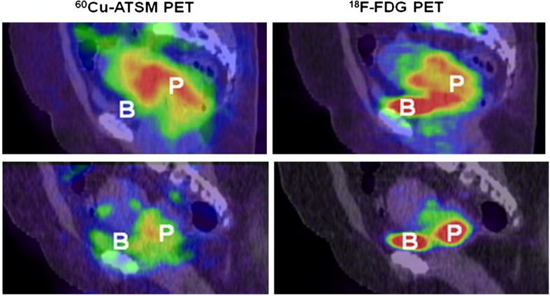
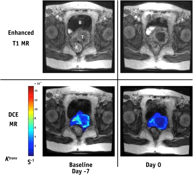
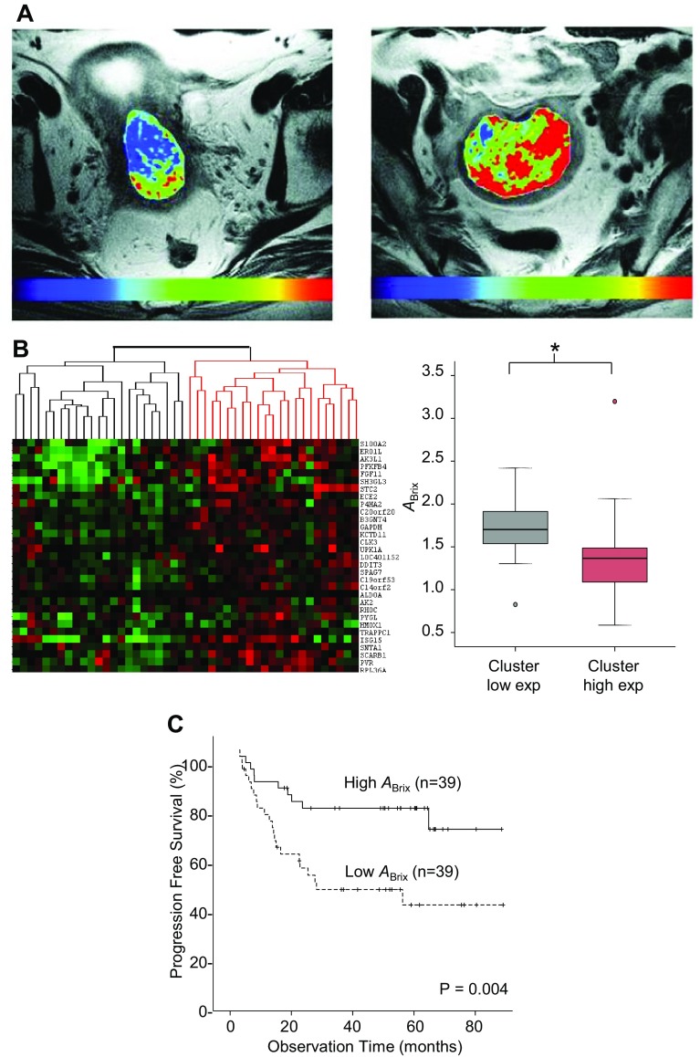
Only a few and rather small studies have utilized PET with tracers specific for hypoxia, and no firm conclusions regarding preferred tracer or clinical potential can be drawn so far. Most studies address indirect hypoxia imaging with dynamic contrast-enhanced techniques. Strong evidences for a role of these techniques in hypoxia imaging have been presented. Pre-treatment images have shown significant association to outcome in several studies, and images acquired during fractionated radiotherapy may further improve risk stratification. Multiparametric MRI and multimodality PET/MRI enable combined imaging of factors of relevance for tumor hypoxia and warrant further investigation.
Several imaging approaches have shown promise for hypoxia imaging in cervical cancer. Evaluation in large clinical trials is required to decide upon the optimal modality and approach.
缺氧成像可能有助于更好地识别有治疗失败风险的宫颈癌患者,并用于治疗规划和监测,但其临床潜力远未得到充分发挥。在此,我们简要描述与成像相关的宫颈肿瘤缺氧生物学,并评估在临床环境中显示出评估缺氧前景的正电子发射断层扫描(PET)和磁共振成像(MRI)技术。我们还将讨论新兴的成像方法,以及成像如何在未来针对缺氧的治疗策略中发挥作用。
我们在PubMed上进行了文献检索,使用与宫颈癌成像和缺氧相关的关键词,特别关注将成像与其他缺氧测量和治疗结果相关联的研究。
只有少数规模较小的研究使用了针对缺氧的特异性示踪剂进行PET检查,目前尚无关于首选示踪剂或临床潜力的确切结论。大多数研究采用动态对比增强技术进行间接缺氧成像。已有充分证据表明这些技术在缺氧成像中发挥作用。在多项研究中,治疗前图像已显示出与治疗结果有显著关联,而在分次放疗期间获取的图像可能进一步改善风险分层。多参数MRI和多模态PET/MRI能够对与肿瘤缺氧相关的因素进行联合成像,值得进一步研究。
几种成像方法在宫颈癌缺氧成像方面已显示出前景。需要通过大型临床试验进行评估,以确定最佳模式和方法。