Department of Immunology, Lerner Research Institute, Cleveland, USA.
Institutes of Head and Neck, Dermatology and Plastic Surgery, Cleveland, USA.
J Immunother Cancer. 2017 Aug 15;5(1):65. doi: 10.1186/s40425-017-0269-7.
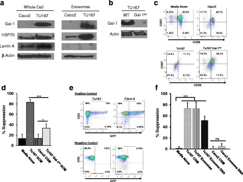
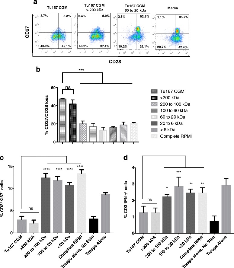
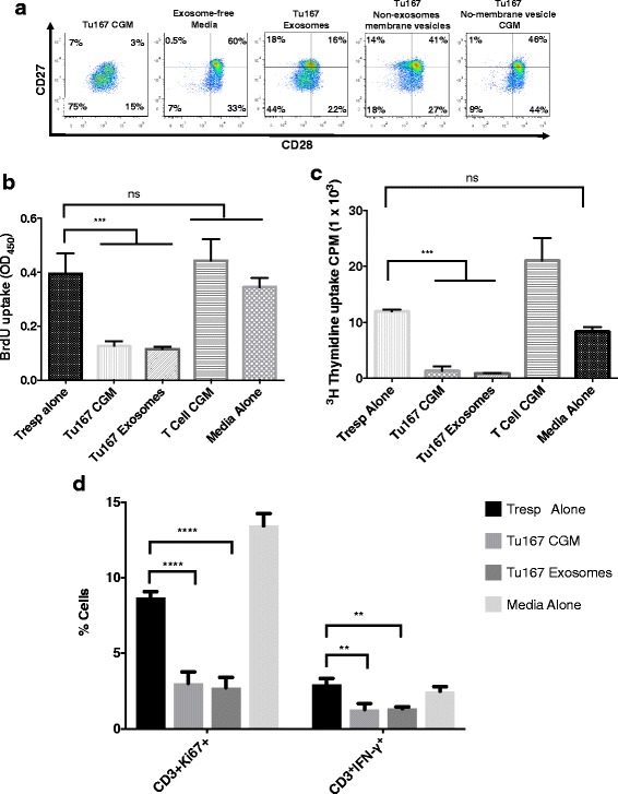
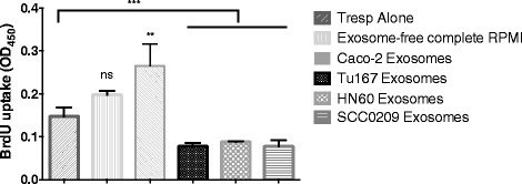
The suppressive nature of immune cells in the tumor microenvironment plays a major role in regulating anti-tumor immune responses. Our previous work demonstrated that a soluble factor from tumor cells is able to induce a suppressor phenotype (SP) in human CD8 T cells typified by loss of CD27/CD28 expression and acquisition of a potent suppressor function. The present study hypothesized that the soluble mechanism that is inducing the SP in CD8 T cells are tumor-derived exosomes (TDEs).
Membrane vesicles and TDEs from multiple head and neck cancer cell line's conditioned growth media were isolated by ultracentrifugation and precipitation, respectively. Human purified CD3CD8 T cells were assessed for their induction of the T cell SP by flow cytometry identifying loss of CD27/CD28 expression and in vitro suppression assays. Furthermore, the T cell SP was characterized for the attenuation of IFN-γ production. To delineate exosomal proteins contributing to T cell SP, mass spectrometry was used to identify unique proteins that were present in TDEs. CRISPR/Cas9 knockout constructs were used to examine the role of one of these proteins, galectin-1. To assess the role of exosomal RNA, RNA purified from TDEs was nucleofected into CD8 T cells followed by suppression analysis.
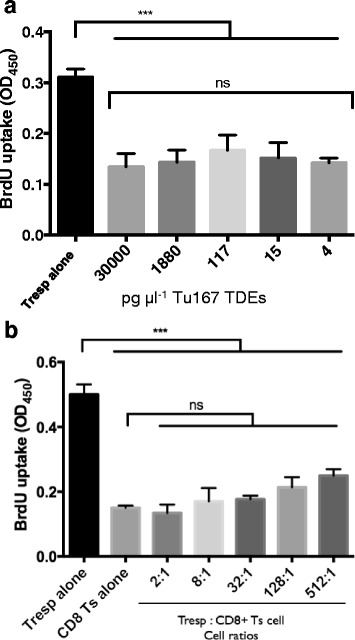
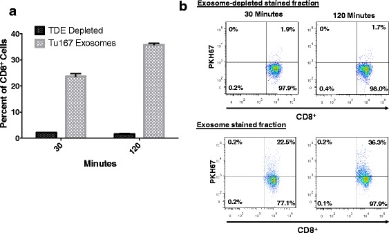
Using fractionated conditioned growth media, factors >200 kDa induced CD8 T cell SP, which was determined to be an exosome by mass spectrometry analysis. Multiple head and neck cancer-derived cell lines were found to secrete T cell SP-inducing exosomes. Mass spectrometry analysis revealed that an immunoregulatory protein, galectin-1 (Gal-1), was expressed in those exosomes, but not in TDEs unable to induce T cell SP. Galectin-1 knockout cells were found to be less able to induce T cell SP. Furthermore, RNA purified from the T cell SP-inducing exosomes were found to partially induce the SP when transfected into normal CD8 T cells.
For the first-time, TDEs have been identified to induce a SP in CD8 T cells and their mode of action may be synergistic effects from exosomal proteins and RNA. One protein in particular, galectin-1, appears to play a significant role in inducing T cell SP. Therefore, tumor-derived immunosuppressive exosomes are a potential therapeutic target to prevent T cell dysfunction and enhance anti-tumor immune responses.
肿瘤微环境中免疫细胞的抑制作用在调节抗肿瘤免疫反应中起着重要作用。我们之前的工作表明,肿瘤细胞中的一种可溶性因子能够诱导人 CD8 T 细胞出现抑制性表型(SP),其特征是丧失 CD27/CD28 表达并获得强大的抑制功能。本研究假设诱导 CD8 T 细胞 SP 的可溶性机制是肿瘤衍生的外泌体(TDE)。
通过超速离心和沉淀分别从多个头颈部癌细胞系的条件培养基中分离出膜囊泡和 TDE。通过流式细胞术鉴定人纯化的 CD3+CD8+T 细胞诱导 T 细胞 SP 的情况,鉴定 CD27/CD28 表达缺失和体外抑制试验。此外,还对 T 细胞 SP 进行了特征分析,以确定其 IFN-γ产生的衰减情况。为了阐明参与 T 细胞 SP 的外泌体蛋白,采用质谱技术鉴定了 TDE 中存在的独特蛋白。使用 CRISPR/Cas9 敲除构建体研究了其中一种蛋白质,半乳糖凝集素-1(Galectin-1)的作用。为了评估外泌体 RNA 的作用,将 TDE 中的 RNA 纯化后进行核转染,然后进行抑制分析。
使用分段条件培养基,>200 kDa 的因子诱导 CD8 T 细胞 SP,通过质谱分析确定为外泌体。发现多个头颈部癌症衍生细胞系分泌诱导 T 细胞 SP 的外泌体。质谱分析表明,一种免疫调节蛋白,半乳糖凝集素-1(Gal-1),在这些外泌体中表达,但不在不能诱导 T 细胞 SP 的 TDE 中表达。Gal-1 敲除细胞被发现诱导 T 细胞 SP 的能力降低。此外,从诱导 T 细胞 SP 的外泌体中纯化的 RNA 在转染到正常 CD8 T 细胞时被发现部分诱导 SP。
本研究首次鉴定出 TDE 可诱导 CD8 T 细胞出现 SP,其作用模式可能是外泌体蛋白和 RNA 的协同作用。特别是一种蛋白质,半乳糖凝集素-1(Gal-1),似乎在诱导 T 细胞 SP 中起着重要作用。因此,肿瘤衍生的免疫抑制性外泌体是预防 T 细胞功能障碍和增强抗肿瘤免疫反应的潜在治疗靶点。