Rathcke Martin, Tranum-Jensen Jørgen, Krogsgaard Michael Rindom
Martin Rathcke, Michael Rindom Krogsgaard, Section for Sportstraumatology M51, Bispebjerg-Frederiksberg Hospital, DK-2400 Copenhagen NV, Denmark.
World J Orthop. 2017 Jul 18;8(7):536-544. doi: 10.5312/wjo.v8.i7.536.
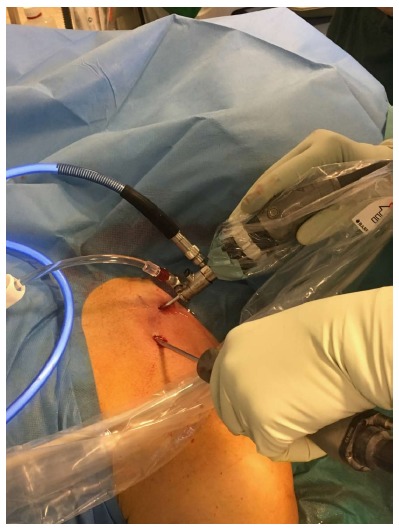
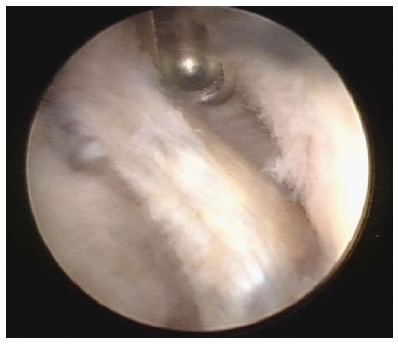
To investigate if there are typical degenerative changes in the ageing sternoclavicular joint (SCJ), potentially accessible for arthroscopic intervention.
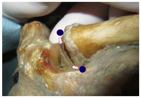
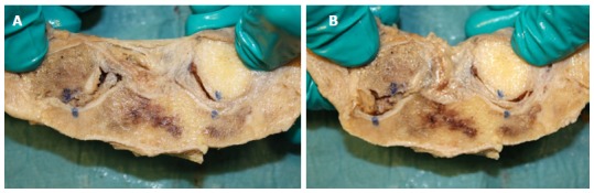
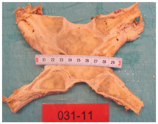
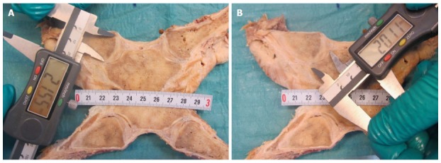
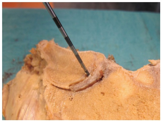
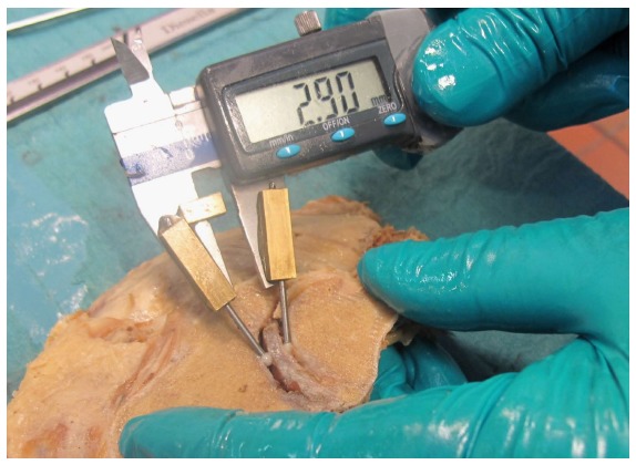
Both SCJs were obtained from 39 human cadavers (mean age: 79 years, range: 59-96, 13 F/26 M). Each frozen specimen was divided frontally with a band saw, so that both SCJs were opened in the same section through the center of the discs. After thawing of the specimens, the condition of the discs was evaluated by probing and visual inspection. The articular cartilages were graded according to Outerbridge, and disc attachments were probed. Cranio-caudal heights of the joint cartilages were measured. Superior motion of the clavicle with inferior movement of the lateral clavicle was measured.
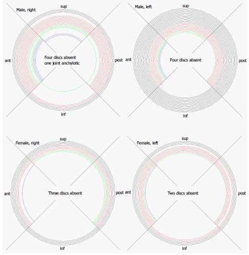
Degenerative changes of the discs were common. Only 22 discs (28%) were fully attached and the discs were thickest superiorly. We found a typical pattern: Detachment of the disc inferiorly in connection with thinning, fraying and fragmentation of the inferior part of the disc, and detachment from the anterior and/or posterior capsule. Severe joint cartilage degeneration ≥ grade 3 was more common on the clavicular side (73%) than on the sternal side (54%) of the joint. In cadavers < 70 years 75% had ≤ grade 2 changes while this was the case for only 19% aged 90 years or more. There was no difference in cartilage changes when right and left sides were compared, and no difference between sexes. Only one cadaver - a woman aged 60 years - had normal cartilages.
Changes in the disc and cartilages can be treated by resection of disc, cartilage, intraarticular osteophytes or medial clavicle end. Reattachment of a degenerated disc is not possible.
研究衰老的胸锁关节(SCJ)是否存在典型的退行性改变,以及这些改变是否适合进行关节镜干预。
从39具人类尸体(平均年龄:79岁,范围:59 - 96岁,13名女性/26名男性)获取双侧胸锁关节。每个冷冻标本用带锯从前向后分割,使双侧胸锁关节在同一截面通过椎间盘中心打开。标本解冻后,通过探查和肉眼检查评估椎间盘状况。根据Outerbridge分级法对关节软骨进行分级,并探查椎间盘附着情况。测量关节软骨的颅尾高度。测量锁骨上移时外侧锁骨下移的情况。
椎间盘退变很常见。只有22个椎间盘(28%)完全附着,且椎间盘最厚处位于上方。我们发现一种典型模式:椎间盘下方分离,同时伴有椎间盘下部变薄、磨损和碎裂,以及与前和/或后关节囊分离。关节软骨严重退变≥3级在关节的锁骨侧(73%)比胸骨侧(54%)更常见。在年龄<70岁的尸体中,75%的软骨改变≤2级,而在90岁及以上的尸体中这一比例仅为19%。左右两侧软骨改变无差异,性别之间也无差异。只有一具尸体——一名60岁女性——软骨正常。
椎间盘和软骨的改变可通过切除椎间盘、软骨、关节内骨赘或锁骨内侧端来治疗。退变的椎间盘无法重新附着。