• 文献检索
  • 文档翻译
  • 深度研究
  • 学术资讯
  • Suppr Zotero 插件Zotero 插件
  • 邀请有礼
  • 套餐&价格
  • 历史记录
应用&插件
Suppr Zotero 插件Zotero 插件浏览器插件Mac 客户端Windows 客户端微信小程序
定价
高级版会员购买积分包购买API积分包
服务
文献检索文档翻译深度研究API 文档MCP 服务
关于我们
关于 Suppr公司介绍联系我们用户协议隐私条款
关注我们

Suppr 超能文献

核心技术专利:CN118964589B侵权必究
粤ICP备2023148730 号-1Suppr @ 2026

文献检索

告别复杂PubMed语法,用中文像聊天一样搜索,搜遍4000万医学文献。AI智能推荐,让科研检索更轻松。

立即免费搜索

文件翻译

保留排版,准确专业,支持PDF/Word/PPT等文件格式,支持 12+语言互译。

免费翻译文档

深度研究

AI帮你快速写综述,25分钟生成高质量综述,智能提取关键信息,辅助科研写作。

立即免费体验

采用超顺磁性氧化铁 MRI 和峰值组织应力探讨腹主动脉瘤的生物学和力学特性:一项联合临床和有限元研究。

Exploring the Biological and Mechanical Properties of Abdominal Aortic Aneurysms Using USPIO MRI and Peak Tissue Stress: A Combined Clinical and Finite Element Study.

机构信息

Centre for Cardiovascular Science, The University of Edinburgh, Edinburgh, UK.

School of Clinical Sciences, The University of Edinburgh, Edinburgh, UK.

出版信息

J Cardiovasc Transl Res. 2017 Dec;10(5-6):489-498. doi: 10.1007/s12265-017-9766-9. Epub 2017 Aug 14.

DOI:10.1007/s12265-017-9766-9
PMID:28808955
原文链接:https://pmc.ncbi.nlm.nih.gov/articles/PMC5722953/
Abstract

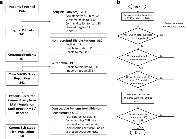
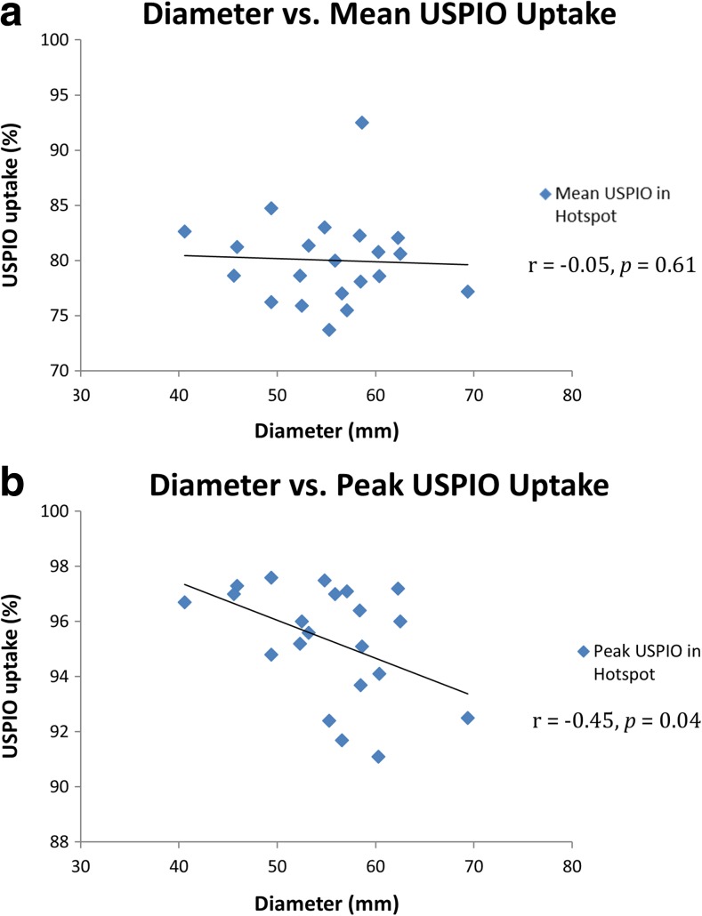
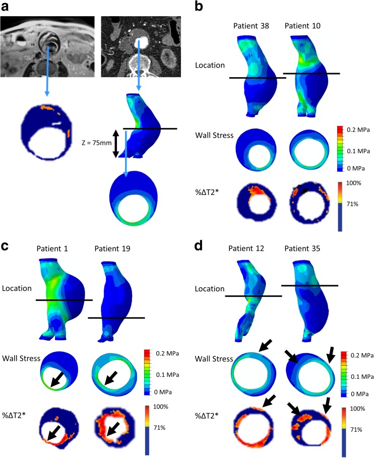
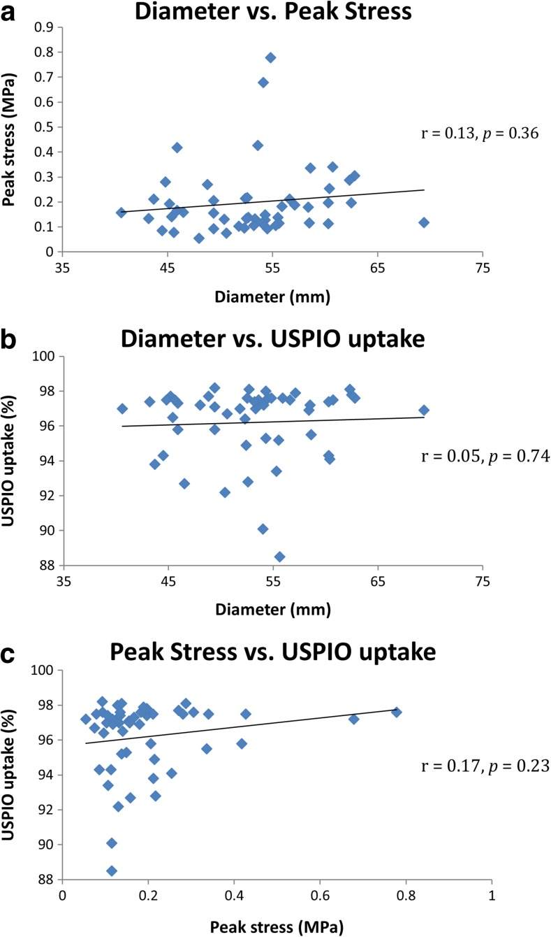
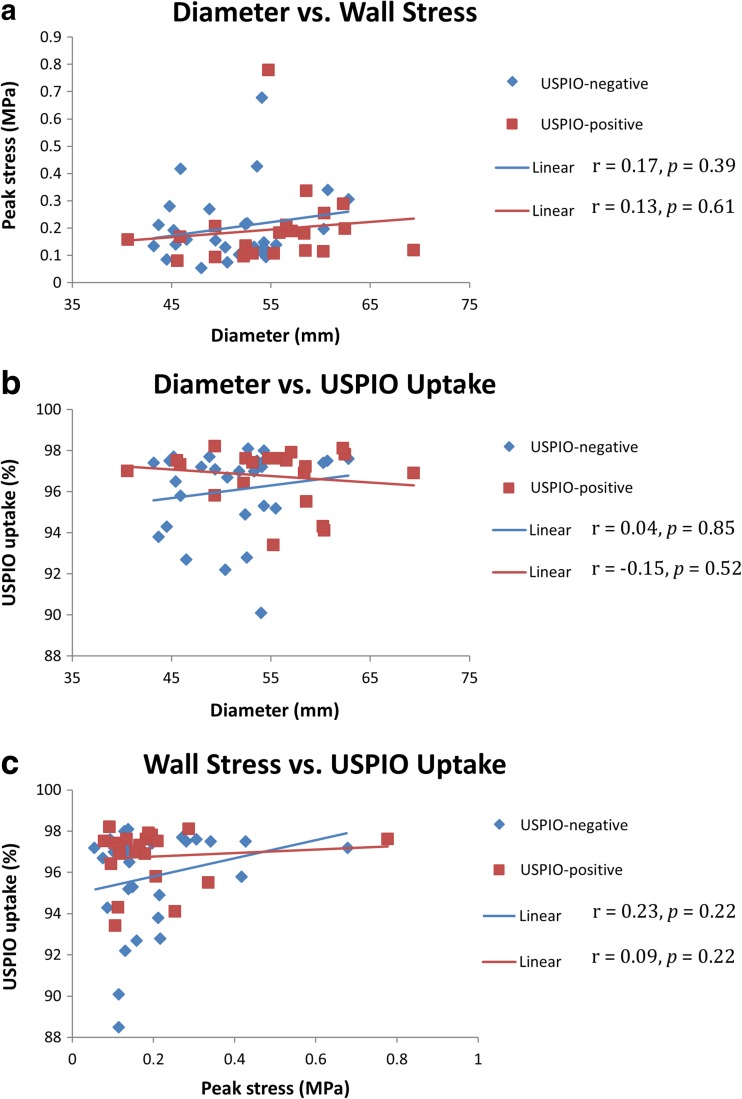
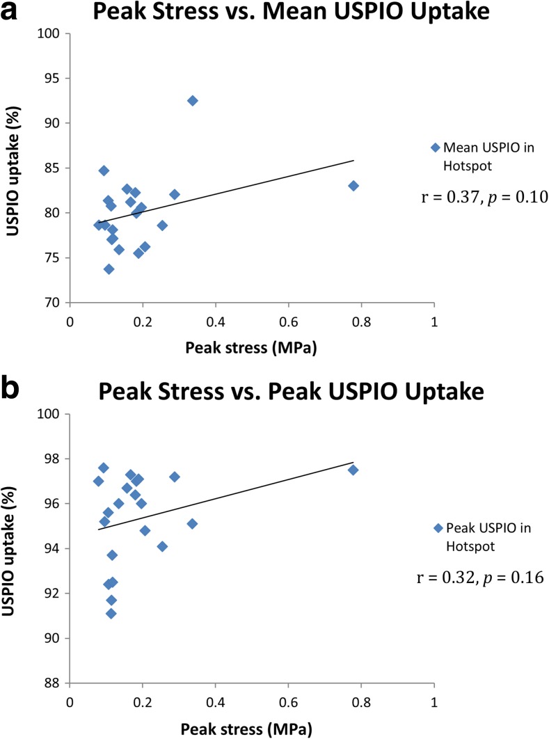
Inflammation detected through the uptake of ultrasmall superparamagnetic particles of iron oxide (USPIO) on magnetic resonance imaging (MRI) and finite element (FE) modelling of tissue stress both hold potential in the assessment of abdominal aortic aneurysm (AAA) rupture risk. This study aimed to examine the spatial relationship between these two biomarkers. Patients (n = 50) > 40 years with AAA maximum diameters > = 40 mm underwent USPIO-enhanced MRI and computed tomography angiogram (CTA). USPIO uptake was compared with wall stress predictions from CTA-based patient-specific FE models of each aneurysm. Elevated stress was commonly observed in areas vulnerable to rupture (e.g. posterior wall and shoulder). Only 16% of aneurysms exhibited co-localisation of elevated stress and mural USPIO enhancement. Globally, no correlation was observed between stress and other measures of USPIO uptake (i.e. mean or peak). It is suggested that cellular inflammation and stress may represent different but complimentary aspects of AAA disease progression.

摘要

在磁共振成像 (MRI) 上检测到的超小超顺磁性氧化铁颗粒 (USPIO) 的摄取和组织应力的有限元 (FE) 建模都有可能用于评估腹主动脉瘤 (AAA) 破裂风险。本研究旨在检查这两种生物标志物之间的空间关系。接受 MRI 和计算机断层血管造影 (CTA) 检查的患者(n=50)年龄>40 岁,AAA 最大直径> =40mm。比较 USPIO 摄取与每个动脉瘤的 CTA 基于患者特定的 FE 模型预测的壁应力。在易破裂的区域(如后壁和肩部)通常观察到升高的应力。只有 16%的动脉瘤显示出升高的壁应力和壁内 USPIO 增强的共定位。总体而言,未观察到应力与 USPIO 摄取的其他测量值(即平均或峰值)之间存在相关性。因此,细胞炎症和应激可能代表 AAA 疾病进展的不同但互补的方面。

https://cdn.ncbi.nlm.nih.gov/pmc/blobs/0a2c/5722953/306b530ec8ba/12265_2017_9766_Fig6_HTML.jpg
https://cdn.ncbi.nlm.nih.gov/pmc/blobs/0a2c/5722953/2e03d4ba3f36/12265_2017_9766_Fig1_HTML.jpg
https://cdn.ncbi.nlm.nih.gov/pmc/blobs/0a2c/5722953/4774525eef2f/12265_2017_9766_Fig2_HTML.jpg
https://cdn.ncbi.nlm.nih.gov/pmc/blobs/0a2c/5722953/73161c2e0430/12265_2017_9766_Fig3_HTML.jpg
https://cdn.ncbi.nlm.nih.gov/pmc/blobs/0a2c/5722953/2b0b5589e2c0/12265_2017_9766_Fig4_HTML.jpg
https://cdn.ncbi.nlm.nih.gov/pmc/blobs/0a2c/5722953/57d452b9e00c/12265_2017_9766_Fig5_HTML.jpg
https://cdn.ncbi.nlm.nih.gov/pmc/blobs/0a2c/5722953/306b530ec8ba/12265_2017_9766_Fig6_HTML.jpg

相似文献

1
Exploring the Biological and Mechanical Properties of Abdominal Aortic Aneurysms Using USPIO MRI and Peak Tissue Stress: A Combined Clinical and Finite Element Study.采用超顺磁性氧化铁 MRI 和峰值组织应力探讨腹主动脉瘤的生物学和力学特性:一项联合临床和有限元研究。
J Cardiovasc Transl Res. 2017 Dec;10(5-6):489-498. doi: 10.1007/s12265-017-9766-9. Epub 2017 Aug 14.
2
Inflammation as a Predictor of Abdominal Aortic Aneurysm Growth and Rupture: A Systematic Review of Imaging Biomarkers.炎症作为腹主动脉瘤生长和破裂的预测指标:影像学生物标志物的系统评价
Eur J Vasc Endovasc Surg. 2016 Sep;52(3):333-42. doi: 10.1016/j.ejvs.2016.05.002. Epub 2016 Jun 6.
3
High Structural Stress and Presence of Intraluminal Thrombus Predict Abdominal Aortic Aneurysm 18F-FDG Uptake: Insights From Biomechanics.高结构应力和腔内血栓的存在可预测腹主动脉瘤的18F-FDG摄取:来自生物力学的见解
Circ Cardiovasc Imaging. 2016 Nov;9(11):e004656. doi: 10.1161/CIRCIMAGING.116.004656.
4
Positron Emission Tomography and Magnetic Resonance Imaging of Cellular Inflammation in Patients with Abdominal Aortic Aneurysms.腹主动脉瘤患者细胞炎症的正电子发射断层扫描和磁共振成像
Eur J Vasc Endovasc Surg. 2016 Apr;51(4):518-26. doi: 10.1016/j.ejvs.2015.12.018. Epub 2016 Feb 23.
5
Finite element analysis in symptomatic and asymptomatic abdominal aortic aneurysms for aortic disease risk stratification.有症状和无症状腹主动脉瘤的有限元分析用于主动脉疾病风险分层。
Int Angiol. 2018 Dec;37(6):479-485. doi: 10.23736/S0392-9590.18.03994-9. Epub 2018 Sep 10.
6
Prediction of Rupture Sites in Abdominal Aortic Aneurysms After Finite Element Analysis.有限元分析后腹主动脉瘤破裂部位的预测
J Endovasc Ther. 2016 Feb;23(1):115-20. doi: 10.1177/1526602815612196. Epub 2015 Oct 23.
7
Use of regional mechanical properties of abdominal aortic aneurysms to advance finite element modeling of rupture risk.利用腹主动脉瘤的局部力学特性来推进破裂风险的有限元建模。
J Endovasc Ther. 2012 Feb;19(1):100-14. doi: 10.1583/11-3456.1.
8
Low wall shear stress predominates at sites of abdominal aortic aneurysm rupture.低壁面切应力在腹主动脉瘤破裂部位占主导。
J Vasc Surg. 2016 Jun;63(6):1613-9. doi: 10.1016/j.jvs.2015.01.040. Epub 2015 Mar 7.
9
Morphological and Biomechanical Features in Abdominal Aortic Aneurysm with Long and Short Neck-Case-Control Study in 64 Abdominal Aortic Aneurysms.长颈部和短颈部腹主动脉瘤的形态学和生物力学特征——64例腹主动脉瘤病例对照研究
Ann Vasc Surg. 2017 Nov;45:223-230. doi: 10.1016/j.avsg.2017.06.054. Epub 2017 Jun 28.
10
Finite element analysis of abdominal aortic aneurysms: predicted rupture risk correlates with aortic wall histology in individual patients.腹主动脉瘤的有限元分析:个体患者中预测的破裂风险与主动脉壁组织学相关。
J Endovasc Ther. 2014 Aug;21(4):556-64. doi: 10.1583/14-4695.1.

引用本文的文献

1
The future of cardiovascular magnetic resonance imaging in thoracic aortopathy: blueprint for the paradigm shift to improve management.心血管磁共振成像在胸主动脉病变中的未来:改善管理的范式转变蓝图。
J Cardiovasc Magn Reson. 2025 Feb 21;27(1):101865. doi: 10.1016/j.jocmr.2025.101865.
2
Geometric and biomechanical modeling aided by machine learning improves the prediction of growth and rupture of small abdominal aortic aneurysms.基于机器学习的几何和生物力学建模有助于提高对小型腹主动脉瘤生长和破裂的预测。
Sci Rep. 2021 Sep 10;11(1):18040. doi: 10.1038/s41598-021-96512-3.
3
Nanoparticle-Assisted Diagnosis and Treatment for Abdominal Aortic Aneurysm.

本文引用的文献

1
Aortic Wall Inflammation Predicts Abdominal Aortic Aneurysm Expansion, Rupture, and Need for Surgical Repair.主动脉壁炎症可预测腹主动脉瘤的扩张、破裂及手术修复需求。
Circulation. 2017 Aug 29;136(9):787-797. doi: 10.1161/CIRCULATIONAHA.117.028433. Epub 2017 Jul 18.
2
Patient-specific modelling of abdominal aortic aneurysms: The influence of wall thickness on predicted clinical outcomes.腹主动脉瘤的患者特异性建模:壁厚对预测临床结果的影响。
Med Eng Phys. 2016 Jun;38(6):526-37. doi: 10.1016/j.medengphy.2016.03.003. Epub 2016 Apr 4.
3
Positron Emission Tomography and Magnetic Resonance Imaging of Cellular Inflammation in Patients with Abdominal Aortic Aneurysms.
纳米颗粒辅助诊断与治疗腹主动脉瘤
Front Med (Lausanne). 2021 Jul 7;8:665846. doi: 10.3389/fmed.2021.665846. eCollection 2021.
4
Targeting the Extracellular Matrix in Abdominal Aortic Aneurysms Using Molecular Imaging Insights.运用分子影像学见解靶向治疗腹主动脉瘤的细胞外基质。
Int J Mol Sci. 2021 Mar 7;22(5):2685. doi: 10.3390/ijms22052685.
5
On the Relative Impact of Intraluminal Thrombus Heterogeneity on Abdominal Aortic Aneurysm Mechanics.腔内血栓异质性对腹主动脉瘤力学的相对影响
J Biomech Eng. 2019 Nov 1;141(11):1110101-11101010. doi: 10.1115/1.4044143.
6
Evaluation of the distribution and progression of intraluminal thrombus in abdominal aortic aneurysms using high-resolution MRI.使用高分辨率 MRI 评估腹主动脉瘤管腔内血栓的分布和进展。
J Magn Reson Imaging. 2019 Sep;50(3):994-1001. doi: 10.1002/jmri.26676. Epub 2019 Jan 29.
7
Predictions and insights for the abdominal aortic aneurysm.腹主动脉瘤的预测与见解。
J Thorac Dis. 2018 Nov;10(11):5982-5984. doi: 10.21037/jtd.2018.10.15.
8
Abdominal aortic aneurysms.腹主动脉瘤。
Nat Rev Dis Primers. 2018 Oct 18;4(1):34. doi: 10.1038/s41572-018-0030-7.
腹主动脉瘤患者细胞炎症的正电子发射断层扫描和磁共振成像
Eur J Vasc Endovasc Surg. 2016 Apr;51(4):518-26. doi: 10.1016/j.ejvs.2015.12.018. Epub 2016 Feb 23.
4
Monitoring the biological activity of abdominal aortic aneurysms Beyond Ultrasound.超越超声监测腹主动脉瘤的生物活性
Heart. 2016 Jun 1;102(11):817-24. doi: 10.1136/heartjnl-2015-308779. Epub 2016 Feb 15.
5
A simple, effective and clinically applicable method to compute abdominal aortic aneurysm wall stress.一种计算腹主动脉瘤壁应力的简单、有效且临床适用的方法。
J Mech Behav Biomed Mater. 2016 May;58:139-148. doi: 10.1016/j.jmbbm.2015.07.029. Epub 2015 Aug 5.
6
MRI using ultrasmall superparamagnetic particles of iron oxide in patients under surveillance for abdominal aortic aneurysms to predict rupture or surgical repair: MRI for abdominal aortic aneurysms to predict rupture or surgery-the MA(3)RS study.在接受腹主动脉瘤监测的患者中使用超小型超顺磁性氧化铁进行磁共振成像以预测破裂或手术修复:用于预测腹主动脉瘤破裂或手术的磁共振成像——MA(3)RS研究
Open Heart. 2015 Apr 18;2(1):e000190. doi: 10.1136/openhrt-2014-000190. eCollection 2015.
7
Local Quantification of Wall Thickness and Intraluminal Thrombus Offer Insight into the Mechanical Properties of the Aneurysmal Aorta.血管壁厚度和腔内血栓的局部定量分析有助于深入了解动脉瘤样主动脉的力学特性。
Ann Biomed Eng. 2015 Aug;43(8):1759-71. doi: 10.1007/s10439-014-1222-2. Epub 2015 Jan 29.
8
Determining the influence of calcification on the failure properties of abdominal aortic aneurysm (AAA) tissue.确定钙化对腹主动脉瘤(AAA)组织失效特性的影响。
J Mech Behav Biomed Mater. 2015 Feb;42:154-67. doi: 10.1016/j.jmbbm.2014.11.005. Epub 2014 Nov 11.
9
Correlation of biomechanics to tissue reaction in aortic aneurysms assessed by finite elements and [18F]-fluorodeoxyglucose-PET/CT.通过有限元法和[18F] - 氟脱氧葡萄糖 - PET/CT评估主动脉瘤生物力学与组织反应的相关性。
Int J Numer Method Biomed Eng. 2012 Apr;28(4):456-71. doi: 10.1002/cnm.1477. Epub 2011 Dec 9.
10
Regions of high wall stress can predict the future location of rupture of abdominal aortic aneurysm.
Cardiovasc Intervent Radiol. 2014 Jun;37(3):815-8. doi: 10.1007/s00270-014-0864-7. Epub 2014 Feb 20.